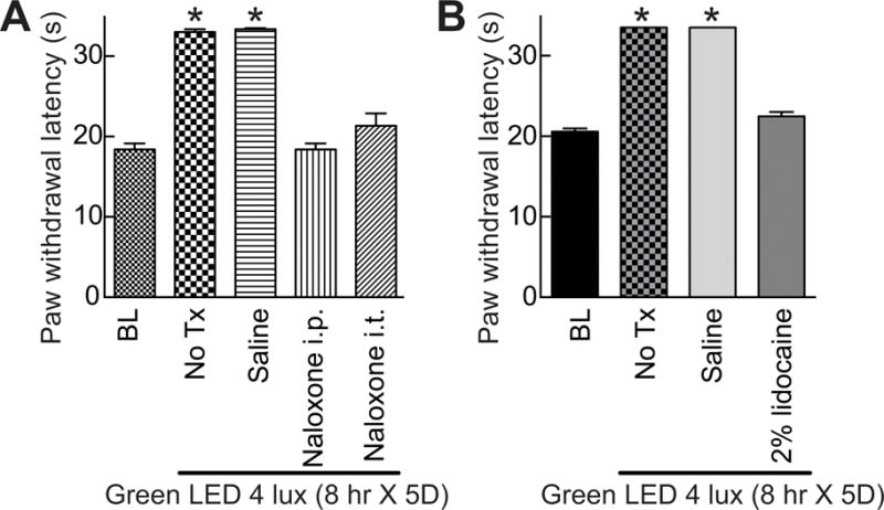
Figure 6. Green light emitting diode (GLED)-induced thermal analgesia involves activity of the descending pain pathways through endogenous opioid signaling.

(A) Bar graph of paw withdrawal latency (seconds) of rats (n=6 per group) prior to and after treatment with GLED as indicated. BL indicates the baseline latency before GLED exposure. Inhibiting mu-opioid receptor (MOR) with naloxone (intraperitoneal (i.p.) or intrathecal (i.t.) administration) (see Table 1) reversed GLED-induced thermal analgesia. Un-treated (No Tx) rats or rats injected i.p. with saline (n=6 per group) developed GLED-induced thermal analgesia. (B) Inactivation of the descending pathway pain with an injection of a 2% solution of lidocaine into the rostral ventromedial medulla (RVM) of rats (n=6 per group) reversed GLED-induced thermal analgesia. *p<0.05 when comparing to BL (one-way ANOVA followed by Student-Newman-Keuls test).
