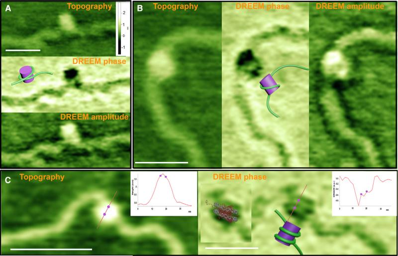
Figure 2. Representative Topographic AFM and DREEM Images of Nucleosomes.
(A and B) Topographic (A top, B left), DREEM-phase (A middle, B center), and DREEM amplitude (A bottom, B right) images of nucleosomes showing one DNA wrapping around histones one time.
(C) Topographic (left) and DREEM-phase (right) images of a nucleosome showing DNA wrapping around nucleosomes twice. Insets show graphs of the height cross-section for the line drawn across the nucleosome in topographic (left) and DREEM-phase (right) images. The two dots on the graph correspond to the positions of the two dots shown on the line across the image, which mark the position of the peaks corresponding to the DNA in the DREEM image. The distance between the two peaks corresponding to the two DNA double strands (dots on graph) is 3.4 nm, which is similar to that seen in the crystal structure (~3 nm) (Luger et al., 1997). Cartoon models of the DNA wrapping around histones are shown on each DREEM-phase image (models are not to scale). The crystal structure of a nucleosome (Luger et al., 1997) overlaid on the DREEM-phase image is shown in the inset of the phase image in (C). The white scale bars are 50 nm. All topographic images are scaled to the same height, and the height scale bar is shown in (A). Both the topographic and DREEM-phase images in (C) are sharper than those in (A) and (B) as a result of a sharper AFM tip. All features in the images are seen in both the trace and retrace scans (Figure S2B). Nucleosomes were reconstituted on a 2,743 bp linear fragment containing 147 bp 601 nucleosome positioning sequence. Unlike the images of nucleosomes, DREEM images of free histones show only smooth “hemispherical shape,” similar to the topographic images (Figure S2A). See also Figure S2.

