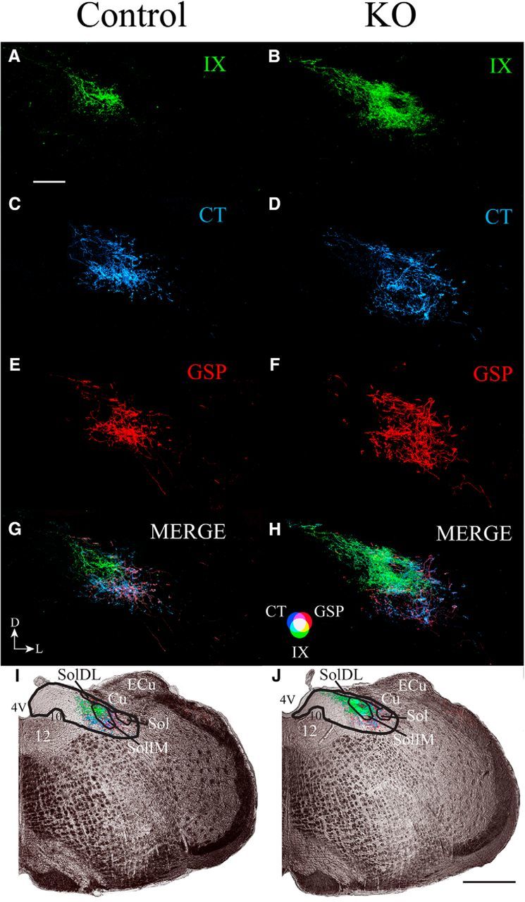
Figure 7.

A–J, Coronal sections through the dorsal/caudal NST showing the IX nerve terminal field (green; A, B), CT nerve terminal field (blue; C, D), GSP nerve terminal field (red; E, F), and merged (G, H) terminal fields, and the terminal fields in the right hemifield of medulla captured with transmitted light (I, J) in control (Control; A, C, E, G, I) and αENaC knock-out (KO; B, D, F, H, J) mice. The orientation of the sections is shown in G. D, Dorsal; L, lateral. The color bar for the merged images in shown in H. Scale bars: A, 200 μm; J, 500 μm. The black lines shown in I and J demarcate the NST (thicker lines) and structures within the NST (thinner lines). 4V, Fourth ventricle; 12, hypoglossal nuclei; 10, dorsal motor nucleus of the vagus; Cu, cuneate nucleus; ECu, external cuneate nucleus; Sol, solitary tract; SolIM, solitary tract nucleus, intermediate; SolDL, solitary tract, dorsolateral. Black, straight lines in I and J point to the relevant structure in the NST.
