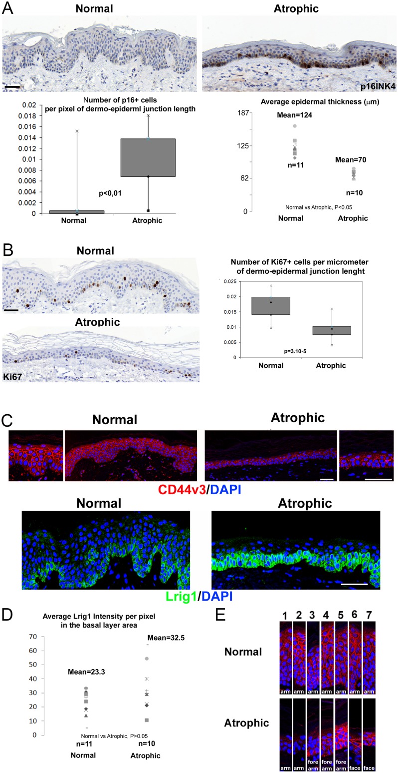
Fig 1. Loss of CD44v3 gradient and retention of Lrig1+ stem cells in senescent atrophic human epidermis.
(A) p16INK4/CDKN2 staining in the epidermis of healthy (n = 11, average age of donors = 32yo with sd = 9) and senescent atrophic (n = 10, average age of donors = 76yo with sd = 13) donors, with quantification of p16INK4/CDKN2 for each group calculated as the number of p16 positive cells divided by the dermal-epidermal junction length and as the average of 3 microscope fields per donor, and the average epidermal thickness of 3 microscope fields per donor, calculated as the epidermal area divided by the length of the dermal-epidermal junction by field. (B) Representative CD44v3 (red) and Lrig1 (green) staining in the epidermis of the normal versus the atrophic group, DNA was counterstained with DAPI (blue). (C) Lrig1 intensity quantification for each donor calculated as the sum of pixel intensities in the green channel (256 levels) in the basal layer standardized to the total basal layer area measured in pixels. (D) CD44v3 staining samples from several individuals of each group, focusing on the CD44v3 expression gradient from the basal layer (down part) to upper differentiated layers (top part). Bar = 55μm.

