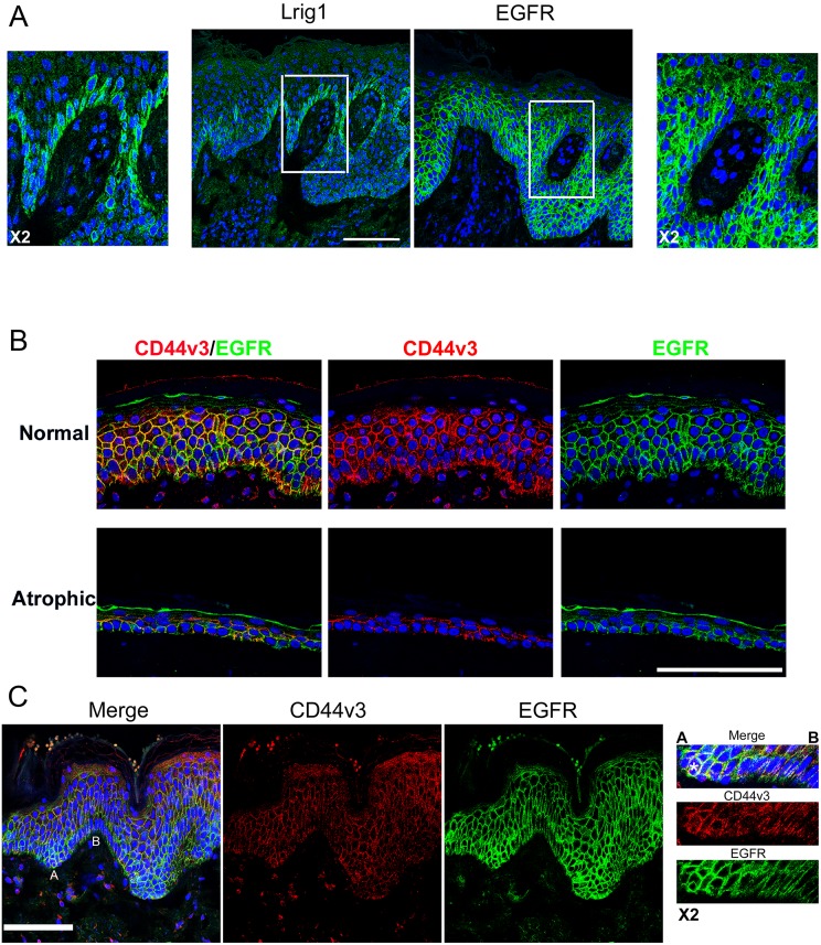
Fig 2. Lrig1 niche lacks EGFR and CD44v3 colocalizes with EGFR.
(A) Lrig1 and EGFR staining (green) in two serial sections of normal human epidermis, blue = DNA stained with DAPI. (B) EGFR (green) and CD44v3 (red) co-staining in healthy and atrophic epidermis, (C) EGFR (green) and CD44v3 (red) co-staining in ridges of healthy epidermis, blue = DNA. Bar = 100μm.

