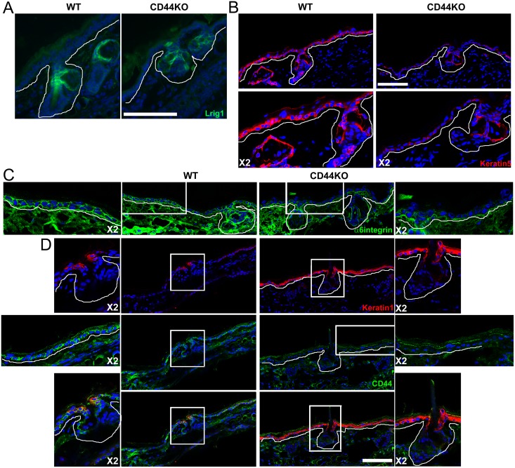
Fig 4. Loss of keratin 5 and α6–integrin expression in the basal layer of the IFE of CD44KO mice.
(A) Lrig1 staining (green), (B) cytokeratin 5 staining (red), (C) α6-integrin staining (green), (D) cytokeratin 1 (red) and CD44 co-staining (green), in the ear epidermis of WT and CD44KO adult mice (>3 months). White line indicates the dermal-epidermal junction. Blue = DAPI. Bar = 87μm.

