1.
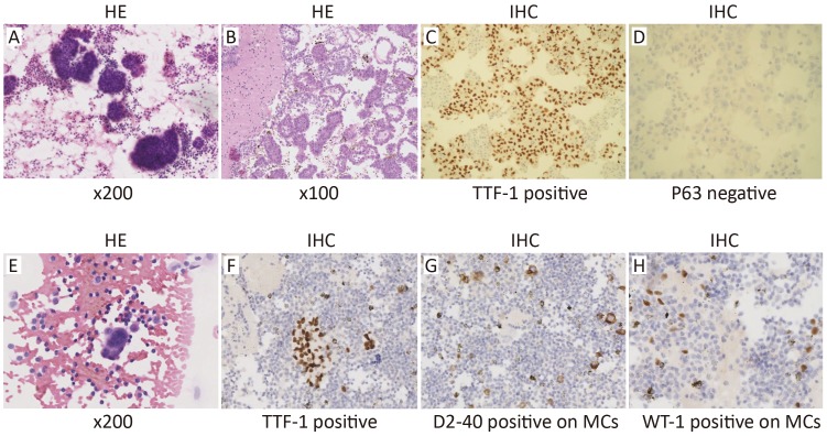
Pathological diagnosis of lung adenocarcinoma with malignant pleural effusion (MPE). (A, E) MPE smears from 2 lung adenocarcinoma patients. (A) is a high tumor content one as numerous cancerous cells are found in the MPE smear, while (E) is a low tumor content one as only a few cancerous cell nests are found; (B) Hematoxylin and eosin (HE) stained section from a high tumor content, formalin-fixed paraffin-embedded (FFPE) MPE cell block (tumor cells >90%); (C, D) In a high tumor content sample, thyroid transcription factor-1 (TTF-1) is diffusely positive (C), and P63 is negative (D); (F) TTF-1-positive signals in sparing focal cancerous cell nests in a low tumor content sample (tumor cells <10%); (G, H) Numerous hyperplastic mesothelial cells (MCs) and inflammatory cells mixed with sparing cancerous cell nests in a low tumor content MPE sample. D2-40 (G) and WT-1 (H) are positive in hyperplastic MCs, but not in cancerous cells in the low tumor content sample.

