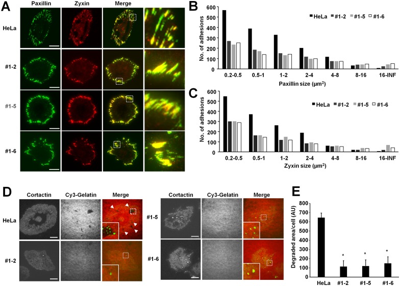
Fig 4. NOX4 knockout reduces focal adhesion and invadopodium formation in HeLa cells.
(A) Cells were seeded in fibronectin coated glass-bottom dishes, and stained with anti-paxillin antibody (green) and anti-zyxin antibody (red). The images were analyzed with TIRF microscope. Bars: 20 μm. (B, C) The area distribution of focal adhesions in 20 cells were analyzed and shown in graph. (D) NOX4 is required for the efficient invadopodium formation. HeLa control and NOX4 knockout cells were cultured on coverslip coated with Cy3-gelatin (red) and stained with Cortactin (green). Gelatin degradation is visualized as darker areas. Arrowheads denote the gelatin degraded areas. Bars: 20 μm. (E) The degradation of gelatin was quantified using image analysis software. The graph shows the average and the standard error of the mean (SEM). HeLa control vs NOX4 knockout cells, *: P<0.05, n = 20.

