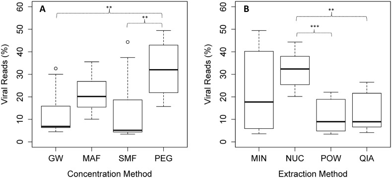
Fig 3. Viral selectivity measured in percentage of reads.
(A) Viral selectivity for the tested concentration methods (B) and extraction methods. Each boxplot was made from 12 individual samples (including the four extraction/concentration methods with three replicates each). The bar, box, whiskers and circles represents median, inter-quartile range, inter-quartile range times 1.5, and outliers, respectively. Asterisks represent significance level of a pairwise t-test with “Holm-Bonferroni” adjusted p-values. ** = p < 0.01, *** = p < 0.001.

