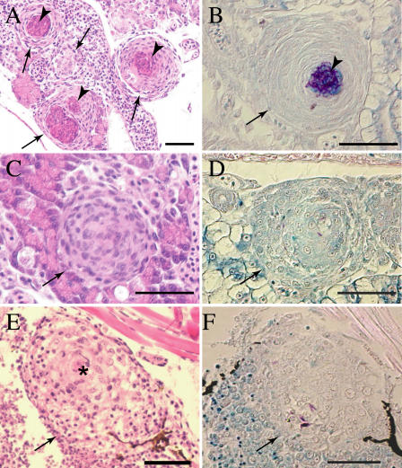
Figure 10. ΔRD1 Infection Is Associated with Persistent Defects in Granuloma Organization.
Tissue histology of 32-d postfertilization fish infected with either 21 WT (A–D) or 9 ΔRD1 (E–F) bacteria (doses were not significantly different p = 0.15) at 1 d postfertilization. Arrows indicate granulomas and loose aggregates, arrowheads indicate caseum. Hematoxylin and eosin staining are shown in (A), (C), and (E), and modified acid-fast staining is shown in (B), (D), and (F). (A) Organized caseating WT granulomas (arrow) with central caseum (arrowhead). (B) WT granuloma showing mycobacteria predominantly in caseum with a few within epithelioid cells. (C) Noncaseating but highly organized WT M. marinum-induced granulomas showing the expected few bacteria within cells in (D). (E) Large, loose, and poorly organized macrophage aggregate of ΔRD1-infected fish with evidence of epithelioid transformation only in the center (denoted by *). (F) A few mycobacteria in the ΔRD1 aggregates. Scale bar, 100 μm. Images in (A–D) were taken with a 40× lens, whereas those in (E) and (F) were taken with a 20× lens.

