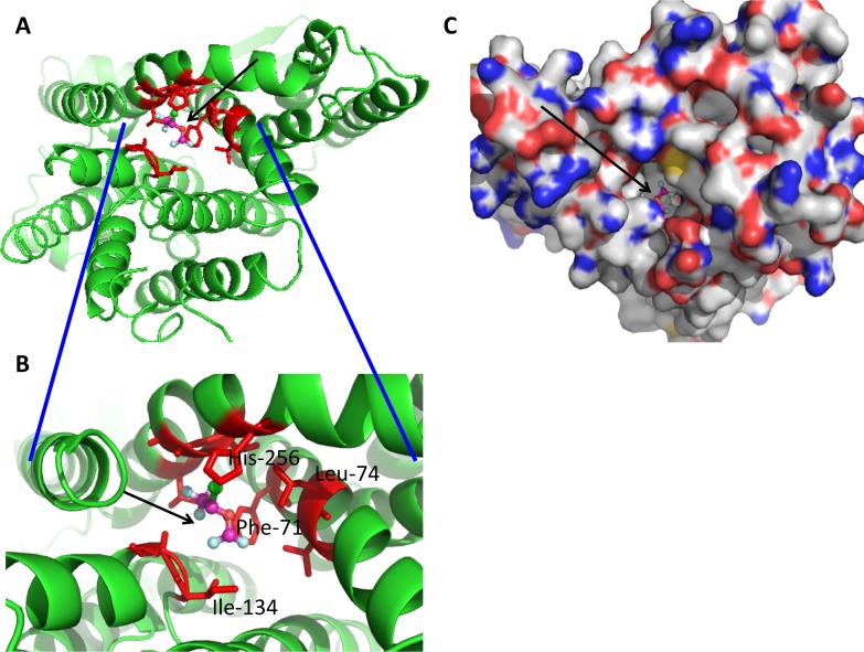
Fig 8. Docking simulation of isoflurane onto NhaA protein (PDB ID: 1ZCD).
Docked image of isoflurane is shown (arrow). In isoflurane, light blue; floride, green; chloride and orange; oxygen. Red residues are within 4 angstroms of isoflurane. (A) Docked image in cartoon. (B) Blowout image of docked site. (C) Docked image in surface.

