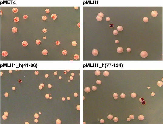
Figure 2.
Appearance of YBT41 colonies following transformation with different MLH1 expression vectors. YBT41 colonies were grown on plates containing low adenine (4 μg/ml) after the introduction of plasmids pMETc, the parental expression vector without an MLH1 gene, pMLH1, pMLH1_h(41-86) and pMLH1_h(77-134) (as indicated). Colonies with red–white sectoring indicate a high level of instability in the ADE2::MS3::ADE2 allele, i.e. mutation to ade2 (mutant cells appear red due to the accumulation of an intermediate in adenine biosynthesis). In all transformations, a background of 10% red colonies was consistently observed and may be a result of mutation in the ADE2::MS3::ADE2 allele prior to, or shortly after, transformation.

