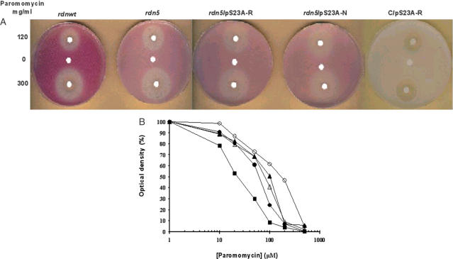
Figure 1.
Inhibition of growth in the presence of paromomycin. (A) Antibiotic disc assay. Cells (∼107) from rdnwt or rdn5 strains were spread on YPD plates and grown at 30°C. Paromomycin at the indicated concentrations or water as control (5 μl) were pipetted onto filter paper discs. Lighter background color shows suppression by rdn5. Clear and white circles around the discs indicate killing and phenotypic suppression, respectively, caused by the antibiotic paromomycin. The C/pS23A-R plate is used as control and it does not contain the ade 1-14 (UGA) marker, therefore it does not indicate suppression; the large clear circles are a measure of sensitivity to paromomycin. (B) Cells were grown in YPD at 30°C to an A660 of 0.9, which was taken as 100%. Concentrations of paromomycin are as indicated. Solid circles, rdnwt strain; solid triangles, rdn5 strain. The other strains are indicated as follows: open circles, rdn5/pS23A-R; open triangles, rdn5/pS23A-N; and solid squares, C/pS23A-R.

