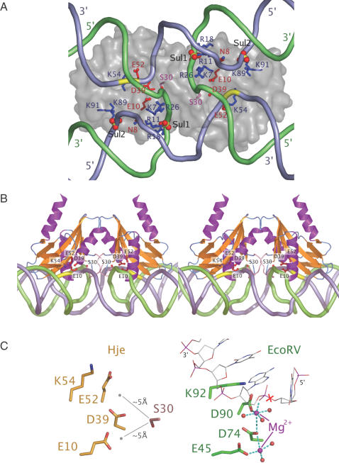
Figure 4.
A model of SsoHje bound to a four-way junction. (A) View along the molecular dyad; semi-transparent protein surface with selected residues shown as ball-and-stick. The scissile phosphate is highlighted in yellow. (B) Stereo view perpendicular to the molecular dyad. (C) Active site residues of Hje compared with an EcoRV–DNA substrate complex [1RVB (39)], indicating the proximity of Ser-30 to the catalytic metal centre.

