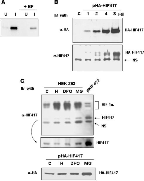
Figure 3.
Generation of a specific antibody against HIF-1α417 and the identification of endogenous HIF-1α417 protein. (A) To identify the specificity of an antibody against HIF-1α417 protein, lysates (50 ng proteins) of IPTG-induced (I) or uninduced (U) bacteria expressing His-tagged HIF-1α417 protein were analyzed by western blotting using anti-HIF-1α417 antibody (α-HIF417). Blocking peptide (BP; 1 μg/ml), which was a bacterially expressed HIF-1α417 protein purified by nickel-affinity chromatography, was added to the primary antibody solution to confirm the antibody specificity. (B) HEK293 cells with various doses of pcDNA–HA-tagged HIF-1α417 (HA-HIF417) using the calcium phosphate method. Total cell lysates (50 μg protein) were analyzed by western blotting using an anti-HA antibody (α-HA) and α-HIF417. NS indicates a non-specific protein cross-reacted with α-HIF417 antiserum. (C) To identify the presence of endogenous HIF-1α417 protein (HIF417), the lysates of untransfected HEK293 cells were analyzed by western blotting using α-HIF417 (upper panel). HEK293 cells were cultured under normoxic (C) or hypoxic (H) conditions, or in the presence of 130 μM desferrioxamine (DFO) or 20 μM MG132 (MG) for 8 h. As a reference for the HIF417 protein, untagged HIF417 expressed in HEK293 cells was also blotted onto the same membrane (pHIF417). The ECL film was developed in a darker shade to compare the HIF417 levels (middle panel). HEK293 cells transfected with pHA-HIF417 were cultured under normoxic (C) or hypoxic (H) conditions, or in the presence of 130 μM desferrioxamine (DFO) or 20 μM MG132 (MG) for 8 h. The cell lysates were analyzed by western blotting using α-HA (lower panel).

