Figure 6.
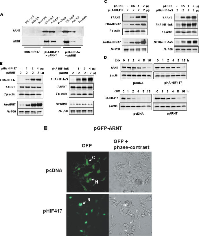
In vivo interaction between HIF-1α417 and ARNT. (A) Association of HIF-1α417 with ARNT. HEK293 cells were co-transfected with pARNT and either pHA-HIF417 or pHA-HIF-1α. Lysates were prepared and immunoprecipitations were performed with α-HA. The co-immunoprecipitation of ARNT with HIF-1α variants was identified by western blotting using α-ARNT. (B) Nuclear translocation of ARNT by HIF-1α417 or full-length HIF-1α. HEK293 cells were co-transfected with pARNT and various doses of pHA-HIF417 (left panel) or pHA-HIF-1αS (right panel). (C) ARNT-dependent expression of HIF-1α417 or full-length HIF-1α. HEK293 cells were co-transfected with various doses of pARNT and 2 μg of pHA-HIF417 (left panel) or pHA-HIF-1αS (right panel). Proteins in total cell lysates (T HIF417, T HIF-1αS, T ARNT, T β-actin) or in nuclear fractions (Nu HIF417, Nu HIF-1αS, Nu ARNT, Nu P50) were analyzed by western blotting using anti-HA and specific antibodies. β-Actin and NF-κB P50 proteins were analyzed as loading controls for total lysates and nuclear extracts, respectively. (D) Stabilization of HIF-1α417 protein by ARNT. HEK293 cells were transfected with pARNT and/or pHA-HIF417, and then treated with 60 μg/ml of cycloheximide (CHX). At the indicated time-points after CHX treatment, the cell lysates were analyzed by western blotting using α-ARNT or α-HA. (E) Nuclear translocation of ARNT by HIF-1α417. The GFP–ARNT expressing plasmid (pGFP–ARNT) was co-transfected into HEK293 cells with pcDNA or pHIF417, and the expression of GFP–ARNT protein was examined by fluorescence microscopy 48 h after transfection. The images in the left panel were captured under a green fluorescence filter, and those in the right panel were overlaid with green fluorescence and phase-contrast images. C, expression in the cytoplasm; N, expression in the nucleus.

