. 2016 Dec 28;5:e19573. doi: 10.7554/eLife.19573

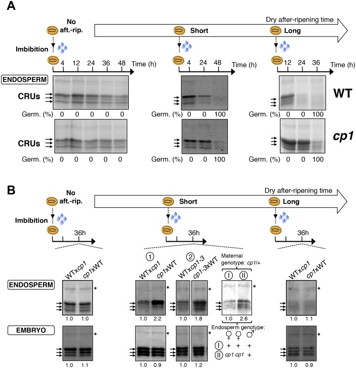
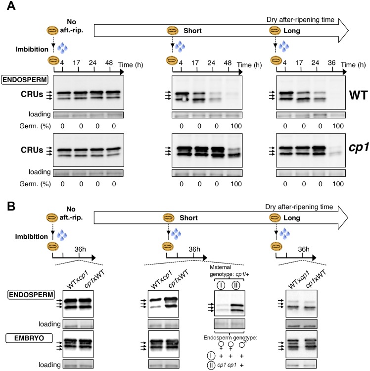
Figure 4. CP1 promotes the decay of CRU proteins.

CRU protein decay is controlled through CP1 maternal gametophytic alleles. (A) Endosperms from non-after-ripened (No aft.-rip.) WT and cp1 seeds or from WT and cp1 seeds after-ripened for short (three days) or long (two months) time periods were dissected at the indicated times after seed imbibition. Proteins isolated from the same number of WT (Col) or cp1 endosperms were separated on SDS PAGE gels and probed with 12S antibody serum. The specificity of the antibody was confirmed using protein extracts isolated from the endosperm and embryo of the cruabc mutant (Figure 4—figure supplement 1). For a given after-ripening time, WT and cp1 endosperm proteins were separated on the same SDS PAGE gel. An aspecific band is used as a loading control. Percent germination at each time-point after seed imbibition is indicated. Arrows indicate highly abundant cruciferin (CRU) proteins. (B) WTxcp1 F1 and cp1xWT F1 seeds (obtained by reciprocally crossing WT (Col) and cp1 plants) were subject to dry after-ripening treatments as in (A). For each after-ripening treatment, endosperms and embryos were dissected 36 hr after seed imbibition. Proteins isolated from the same number of endosperms or embryos were processed as in (A). Seeds obtained after crossing cp1/+ heterozygous mother plants with Col WT (+/+) pollen were after-ripened for a short time period (three days). Endosperms were dissected 36 hr after imbibition, whereas embryos were further cultured for later genotyping in order to distinguish endosperms according to their (i) +/+/+ or (ii) cp1/cp1/+ genotype. Endosperms with the same genotype were pooled. Proteins extracted from the two endosperm pools were processed as in (A).

DOI: http://dx.doi.org/10.7554/eLife.19573.024

Figure 4.

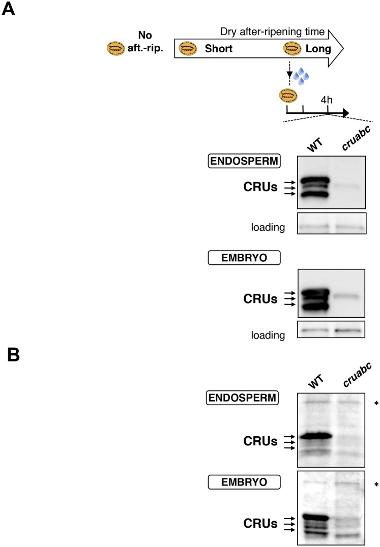
Figure 4—figure supplement 1. Antibody specificity.

Figure 4—figure supplement 1.

(A) Protein gel blot analysis using 12S antibody serum. Protein extracts were isolated from the same number of WT (Col) and cruabc endosperms or embryos harvested 4 hr after seed imbibition. Proteins were separated on SDS PAGE gels. An aspecific band is used as a loading control. (B) Coomassie blue staining of proteins isolated from the same number of WT (Col) and cruabc endosperms or embryos separated on SDS PAGE gels. The asterisk (*) denotes a band used as an internal loading control.
Figure 4—figure supplement 2. CP1 promotes CRU protein decay.

Figure 4—figure supplement 2.

CRU protein decay is controlled through CP1 maternal gametophytic alleles. (A) Endosperms from non-after-ripened (No aft.-rip.) WT and cp1 seeds or from WT and cp1 seeds after-ripened for short (three days) or long (two months) time periods were dissected at the indicated times after seed imbibition. Proteins isolated from the same number of WT (Col) or cp1 endosperms were separated on SDS PAGE gels and stained with coomassie blue. For a given after-ripening time, WT and cp1 endosperm proteins were separated on the same SDS PAGE gel. An aspecific band is used as a loading control. Percent germination at each time-point after seed imbibition is indicated. Arrows indicate highly abundant cruciferin (CRU) proteins. (B) WTxcp1 F1 and cp1xWT F1 seeds (obtained by reciprocally crossing WT (Col) and cp1 plants) were subject to dry after-ripening treatments as in (A). For each after-ripening treatment, endosperms and embryos were dissected 36 hr after seed imbibition. Proteins isolated from the same number of endosperms or embryos were processed as in (A). Seeds obtained after crossing cp1/+ heterozygous mother plants with Col WT (+/+) pollen were after-ripened for a short time period (three days). Endosperms were dissected 36 hr after imbibition, whereas embryos were further cultured for later genotyping in order to distinguish endosperms according to their (i) +/+/+ or (ii) cp1/cp1/+ genotype. Endosperms with the same genotype were pooled. Proteins extracted from the two endosperms pools were processed as in (A). The asterisk (*) denotes a band used as an internal loading control. Numbers indicate protein signal quantification using imageJ software.
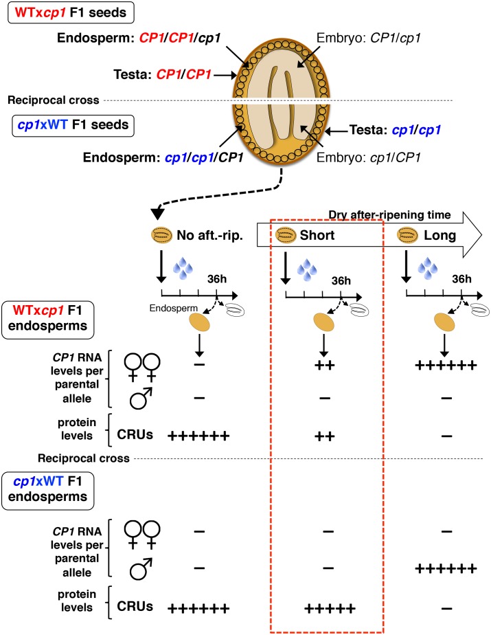
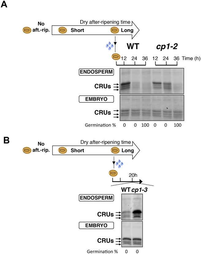
Figure 4—figure supplement 3. CP1 promotes CRU protein decay.

Figure 4—figure supplement 3.

(A) Endosperms from fully after-ripened (two months) WT and cp1-2 (salk _020878) seeds were dissected at the indicated times after seed imbibition. Proteins isolated from the same number of WT (Col) or cp1-2 endosperms were separated on SDS PAGE gels and stained with coomassie blue. The WT samples are the same as those in Figure 4—figure supplement 2. Percent germination at each time-point after seed imbibition is indicated. Arrows indicate highly abundant cruciferin proteins (CRUs). (B) Endosperms from fully after-ripened (two months) WT and cp1-3 (salk _067293) seeds were dissected 20 hr after seed imbibition and processed as in (A).
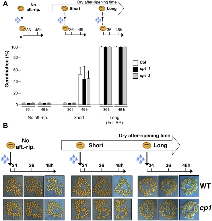
Figure 4—figure supplement 4. CP1 is not required to regulate germination sensu stricto.

Figure 4—figure supplement 4.

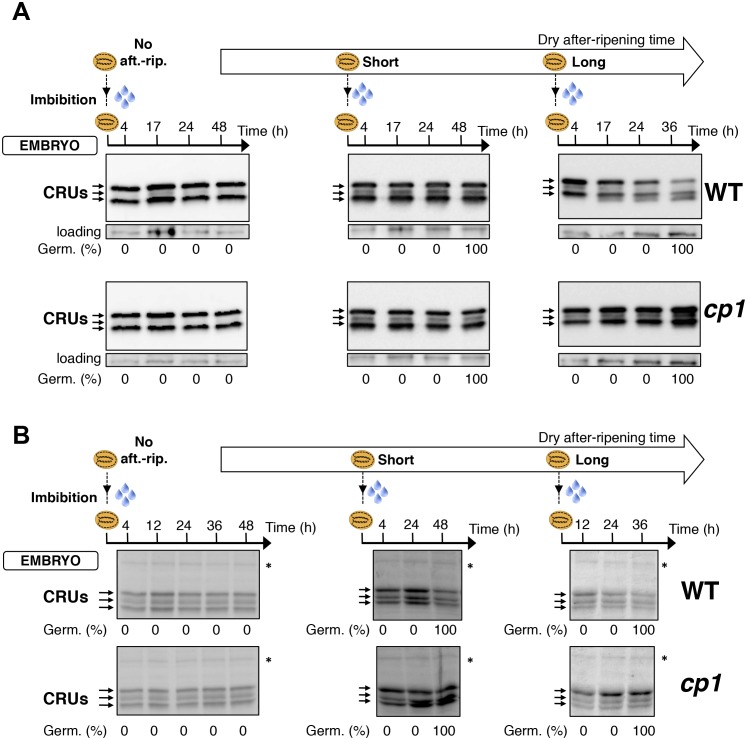
(A) WT (Col), cp1-1 (salk_146500) and cp1-3 (salk_067293) seeds were either not after-ripened (No aft.-rip) or after-ripened for short (three days) or long (two months) time periods. Germination was scored 36 and 48 hr after seed imbibition (4 replicates, n = 150–200). (B) Representative pictures of WT of cp1-1 seeds 24, 36 and 48 hr after seed imbibition. After-ripening times as described in (A).
Figure 4—figure supplement 5. Embryo CRU protein levels are constant during early seed imbibition times.

Figure 4—figure supplement 5.

Embryos from non-after-ripened (No aft.-rip.) WT (Col) and cp1 seeds or from WT and cp1 seeds after-ripened for short (three days) or long (two months) time periods were dissected at the indicated times after seed imbibition. Proteins isolated from the same number of embryos were separated on SDS PAGE gels and probed using 12S antibody serum (A) or stained with coomassie blue (B). For a given after-ripening time, WT and cp1 embryo proteins were separated on the same SDS PAGE gel. Percent germination at each time-point after seed imbibition is indicated. Arrows indicate highly abundant cruciferin proteins (CRUs). An aspecific band is used as a loading control in (A). The asterisk (*) denotes a band used as an internal loading control in (B).
Figure 4—figure supplement 6. Procedure to assess CRUprotein decay through maternal CP1 alleles.

Figure 4—figure supplement 6.

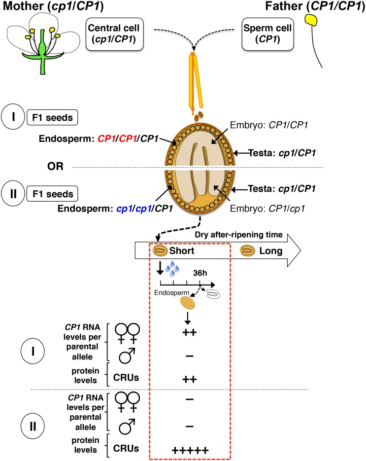
The genotypes of the different tissues present in WTxcp1 F1 and cp1xWT F1 seeds are depicted. In absence of after-ripening, CP1 maternal allele expression is not detectable, which leads to similarly high CRUs protein levels in the WTxcp1 F1 and cp1xWT F1 endosperms upon seed imbibition. In fully after-ripened seeds, CP1 expression is high and biallelic so that both WTxcp1 F1 and cp1xWT F1 endosperms are able to downregulate CRU proteins. By contrast, in shortly after-ripened seeds, preferential CP1 maternal allele expression takes place so that WTxcp1 F1 endosperm cells express higher CP1 mRNA levels than do cp1xWT F1 endosperms. As a result, upon imbibition, WTxcp1 F1 endosperms ought to accumulate lower CRUlevels relative to cp1xWT F1 endosperms.
Figure 4—figure supplement 7. Procedure for the assessment of CRU protein decay through CP1 maternal gametophytic alleles.

Figure 4—figure supplement 7.

Hybrid seeds were obtained after crossing heterozygous cp1/Col mother plants with WT (Col) pollen. The resulting siliques have two types of seeds according to their endosperm tissues: type I bear CP1 maternal genomes, whereas type II bear cp1 maternal genomes. Shortly after-ripened type I seeds are expected to have endosperm tissues that express higher CP1 mRNA levels than do type II seeds. Thus, endosperm from type I seeds should have lower CRU levels relative to that of type II seeds upon seed imbibition.
Figure 4—figure supplement 8. Monoallelic CP1 expression rises upon imbibition in dormant seeds.

Figure 4—figure supplement 8.

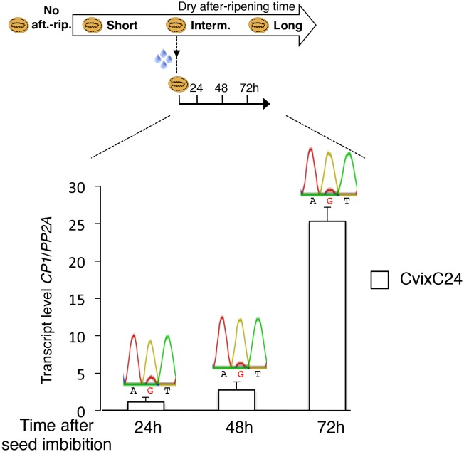
CvixC24 (C24 pollen) F1 seeds were after-ripened for an intermediate time period (25 days). RNA extracted from endosperm 24 hr, 48 hr and 72 hr after seed imbibition, i.e. prior to germination, was used for qPCR and RT-PCR for Sanger sequencing analysis. Expression levels of CP1 were normalized to those of PP2A. CP1 expression levels relative to those found in endosperms dissected at indicated time are shown.