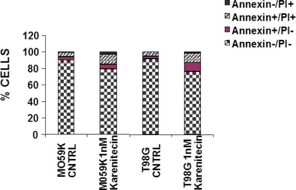
Figure 4.
Karenitecin-induced apoptosis of glioma cell lines as determined by dual colored (PI and Annexin V) flow cytometry. The cell lines were treated with or without 1 nM karenitecin for 72hrs. The cells were harvested by cell dissociation solution washed with PBS and subsequently stained with antibody to annexin V conjugated to FITC and with PI [10 μg/ml]. Viable [annexin V−/PI−] pre-apoptotic [annexin V+/PI−], apoptotic [annexin V+/PI+], and the residual damaged [annexin V−/PI+ cells were quantified by flow cytometric analysis. NOTE: Bar reads top to bottom Annexin−/PI+, Annexin+/PI+, Annexin+/PI−, Annexin−/PI−

