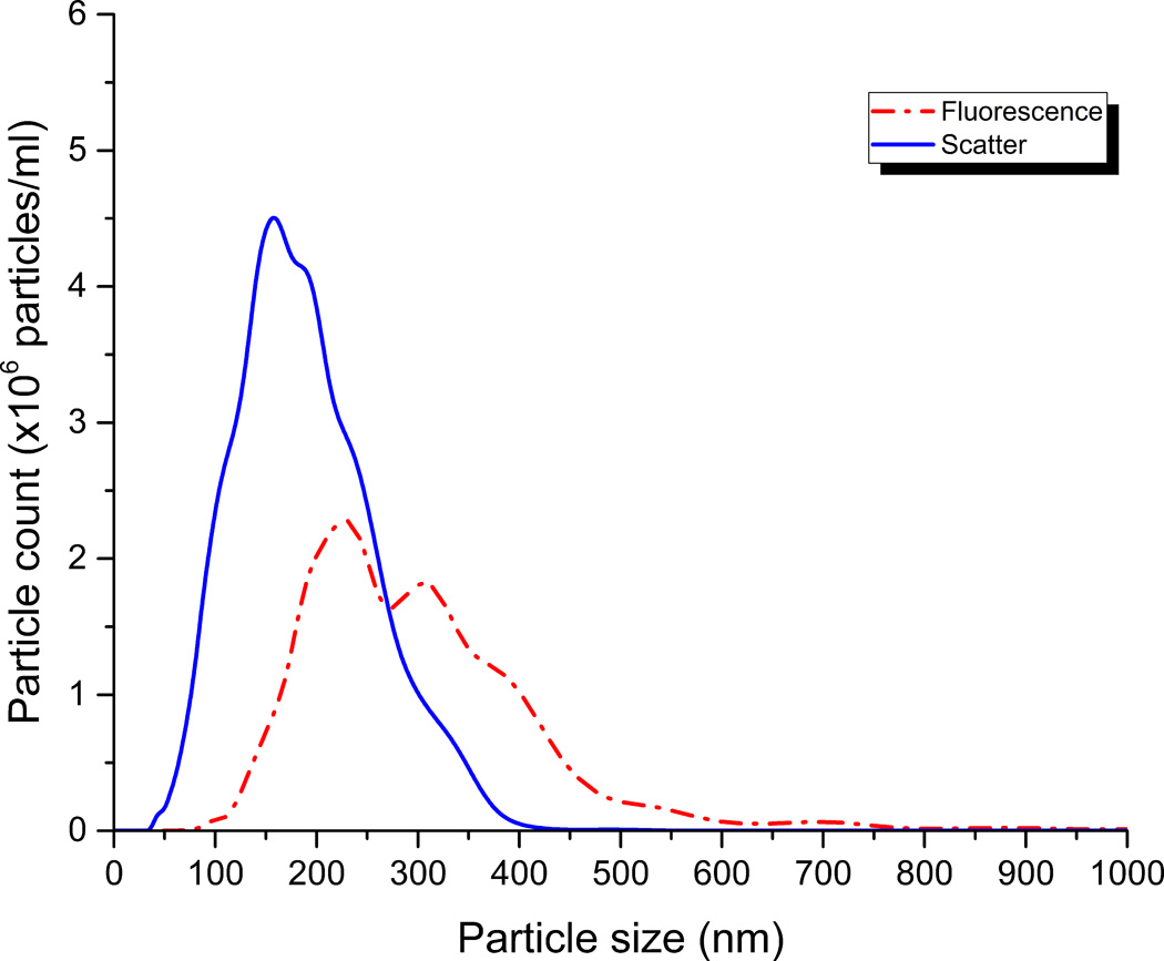
Figure 2. Representative size distributions of EVs and peptide-labeled EVs by nanoparticle tracking analysis.
Alexa 546-labeled MARCKS-ED peptide (110 nM) was incubated with pooled human plasma (1:2000 dilution in HEPES-buffered saline) and analyzed by nanoparticle tracking analysis (NanoSight). Size distributions from representative individual videos taken in scatter mode (blue) to report EV size distribution, and fluorescence mode (red) to report MARCKS-labeled EV size distribution. This illustrates the potential for using fluorescently labeled peptides to probe vesicles from complex ex vivo samples and suggests the presence of a subpopulation of PS+ vesicles.

