
Figure 2. No spontaneous regrowth of CST, AST or 5HT axons after deleting chronic astrocyte scars.
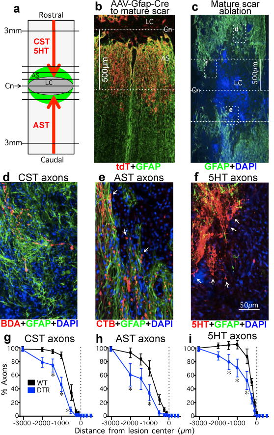
(a) Experiment summary schematic. (b) Selective targeting of tdT reporter to GFAP-positive scar-forming astrocytes in 500μm zone occupied by astrocyte scar (see Extended Data Figure 1c). (c) DTR-DTX mediated ablation of chronic astrocyte scar after severe SCI. Dotted lines indicate Cn and 500μm on either side normally occupied by scar-forming astrocytes in WT mice. Boxes show locations of d–f in adjacent sections. (d) CST axons are found only among GFAP-positive astrocytes proximal to ablated scar. (e) AST axons at margins of large area depleted of scar but have not regrown (arrows). (f) 5HT axons are within area depleted of scar but have not regrown (arrows). (g–i) Numbers of CST (F), AST (G), or 5HT (H) axons at various distances from SCI lesion centers as a percent of the number of axons present at 3mm proximal. n = 6 CST; n = 5 AST and 5HT; * p<0.05 versus WT (ANOVA with Newman-Keuls).