
Figure. 5. Robust regrowth of AST axons can be stimulated after WT SCI and is significantly attenuated by preventing astrocyte scar formation.
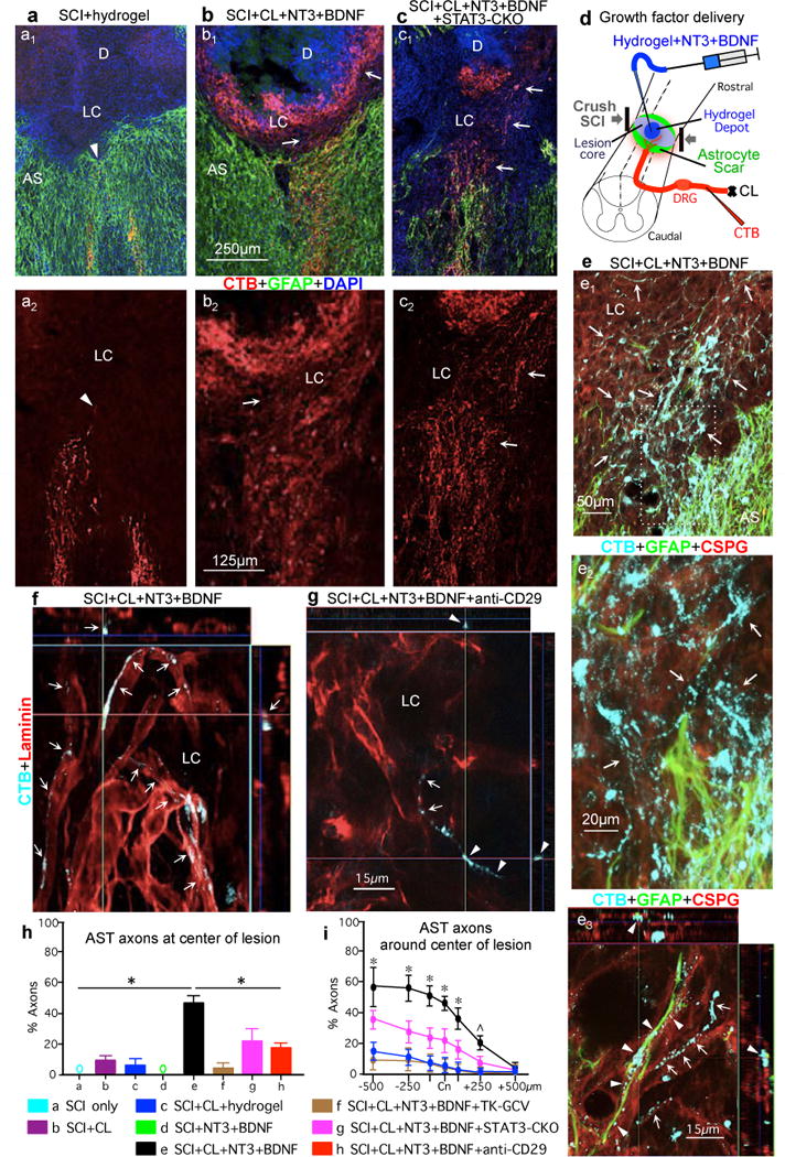
(a1–c1) AST axons (CTB-tracing) plus GFAP immunohistochemistry. (a2–c2) AST axons alone. (a) WT mouse, SCI and hydrogel only (no growth factors). Arrowhead denotes most rostrally penetrating axons that do not pass beyond AS. (b) WT mouse, SCI plus conditioning lesions (CL) and hydrogel depot (D) with NT3+BDNF. Arrows denote robust regrowth of AST axons past AS into LC and along, but not into, the depot that releases NT3+BDNF but provides no adhesive matrix. (c) STAT3-CKO mouse, SCI plus CL and NT3+BDNF depot. Arrows denote regrowth of AST axons into LC. (d) Experiment summary schematic. (e–f) WT mice. (e1–e3) AST plus GFAP and CSPG (CS56) immunohistochemistry. Box in e1 is shown in e2. (e1 and e2) Arrows denote robust regrowth of stimulated AST axons past AS into LC through CSPG. (e3) Regrowing AST axons track along CSPG-positive GFAP-negative structures (arrows) or along CSPG-positive GFAP-positive astrocyte processes (arrowheads) (See Extended Data Figures 7,8). (f,g) AST axons plus laminin immunohistochemistry. (f) Arrows denote regrowing stimulated AST axons tracking along laminin. (g) Arrowheads denote stimulated AST axons exposed to anti-CD29 antibody and failing to maintain contact with laminin. (h,i) Numbers of AST axons at SCI Cn (h) or on either side (i) expressed as percent of axons 3mm proximal. n = 5; * p<0.05 versus all other groups, ˆ p<0.05 versus all groups except STAT3-CKO (ANOVA with Newman-Keuls).