
Extended data figure 4. Comparison of genomic data from astrocytes and non-astrocyte cells from WT and STAT3-CKO mice after SCI.
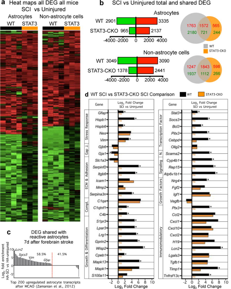
(a) Heat maps depicting all significantly differentially expressed genes (DEG), as determined by RNA-Seq, for WT and STAT3-CKO astrocytes and non-astrocytes from independent biological replicates two weeks after SCI relative to uninjured WT control. Red upregulated, green downregulated. (b) Total numbers and Venn diagrams of significant DEGs in WT and STAT3-CKO astrocytes and non-astrocytes two weeks after SCI relative to uninjured control. Red and green numerical values indicate significantly upregulated and downregulated genes, respectively. (c) Comparison of altered gene expression in our SCI-reactive astrocytes and previously reported forebrain stroke-reactive astrocytes28. Of the 200 most highly elevated genes in forebrain astrocytes 1 week following stroke28, 58.5% (red line) are also significantly elevated in astrocytes after SCI, relative to uninjured. (d) Comparison of expression by WT SCI and STAT3-CKO SCI reactive astrocytes of a selected cross-section of genes that are highly regulated after SCI by WT reactive astrocytes. Many of the regulated genes exhibit changes that are expected and implicated in WT reactive astrogliosis mechanisms and roles, and some of the changes appear to be newly identified in this context. Note that many of the genes are not regulated or exhibit attenuated changes in STAT3-CKO SCI astrocytes. n = 4 for uninjured and WT SCI; n = 3 for STAT3-CKO SCI (SCI-STAT3). FDR<0.1 for differential expression and enrichment analysis.