
Extended data figure 5. Immunohistochemistry of specific CSPGs.
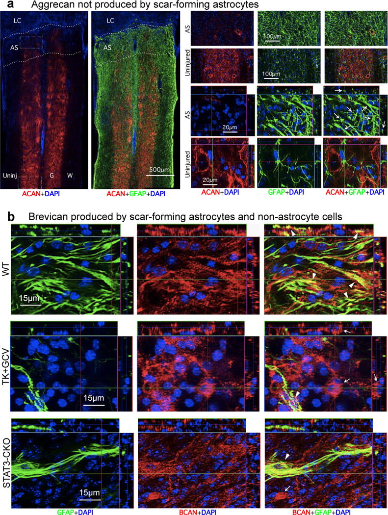
(a) Absence of aggrecan (ACAN) production by scar-forming astrocytes. Images show individual fluorescence channels of ACAN and GFAP immunohistochemistry from horizontal sections two weeks after severe SCI in a representative WT mouse. Boxes denote areas of astrocyte scar (AS) or uninjured tissue (Uninj) shown at higher magnification. Note that ACAN (i) is heavily present in the perineuronal nets that surround neurons in uninjured tissue, (ii) is almost absent from AS and lesion core (LC), and (iii) is not detectably produced by newly generated scar-forming astrocytes (arrows). (b) Brevican (BCAN) production by scar-forming astrocytes and non-astrocyte cells. Images show individual fluorescence channels of BCAN and GFAP immunohistochemistry from horizontal sections two weeks after severe SCI, in WT mice and mice with transgenic ablation (TK+GCV) or attenuation (STAT3-CKO) of astrocyte scar formation. Note that BCAN is produced both by GFAP-positive scar-forming astrocytes (arrowheads) and by non-astrocyte cells (arrows).