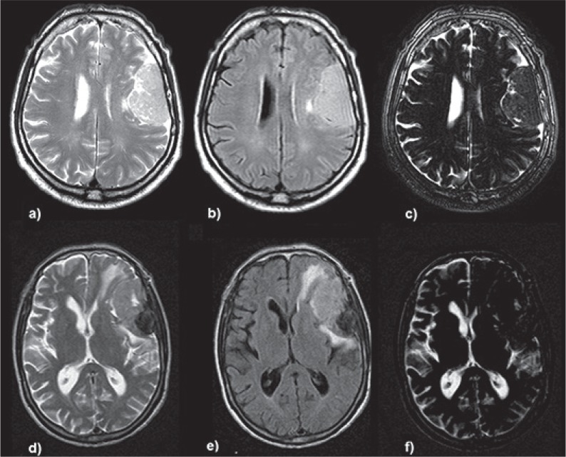
Fig. 1.
CSF sensitive digital image subtraction from T2-FLAIR sequences, two representative cases. Upper row: left parietal convexity meningioma. a) T2 sequence b) FLAIR sequence, c) digital T2-FLAIR subtraction image showing a positive CSF cleft sign with edema suppression, the surgical report describes Simpson 1 resection and good dissection plane on the entire surface of the interface, the patient had no postoperative neurological deficits. Lower row: left fronto-parietal convexity meningioma. d) T2 sequence e) FLAIR sequence f) digital subtraction image showing the lesion with no or minimal CSF cleft sign compared to T2 sequence, also note the peritumoral edema subtraction, the surgical report describes a Simpson 1 resection and bad cleavage plane mainly in its frontal aspect where the tumor was very attached to the cortical surface, the patient presented transient motor aphasia in the immediate postoperative period that fully recovered after 7 days of treatment with steroids and specific speech therapy.

