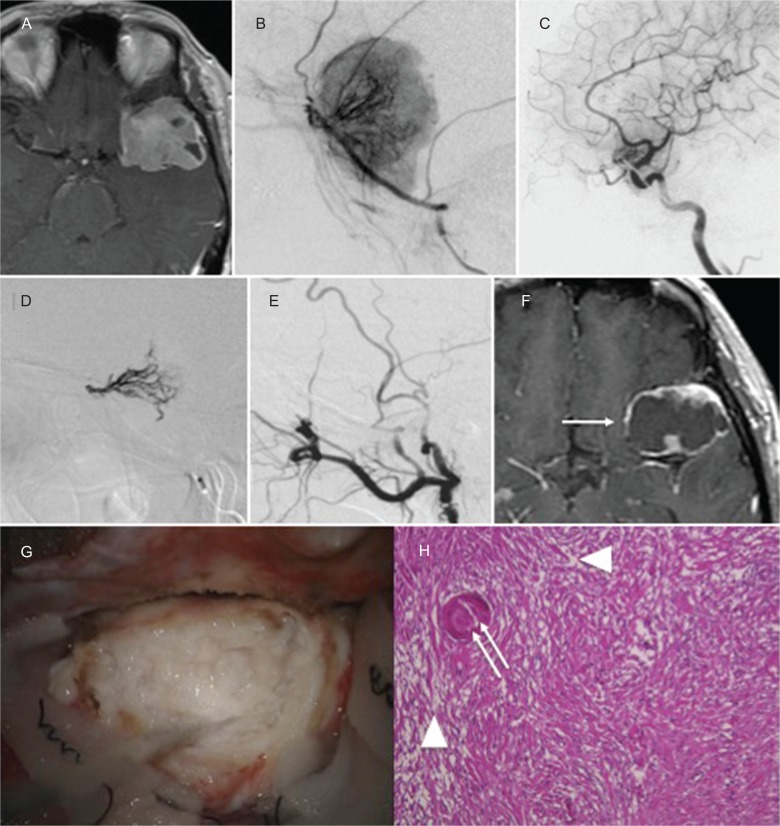
Fig. 3.
(Case 3) Contrast-enhanced T1WI revealed a left sphenoid ridge meningioma (A). Left external carotid angiogram (B) and internal carotid angiogram (C) showed that the tumor was supplied by the left MMA and recurrent meningeal artery from the ophthalmic artery. NBCA was injected into the MMA and recurrent artery from the ophthalmic artery (D). After embolization, the tumor was devascularized (E). MRI taken 3 weeks after embolization demonstrated remarkable shrinkage (F: white arrow) of the tumor. Intraoperative photographs revealed a whitish and degenerated tumor (G). Histopathologic findings (H) showed necrotic changes in the tumor (white arrowhead) and NBCA cast in the tumor vessels (white double arrow).

