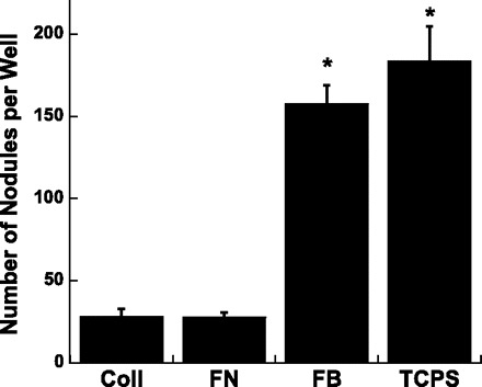
Fig. 1.

The number of calcified nodules formed by valvular interstitial cells (VICs) cultured on different substrate coatings for 5 days; nodules were identified and counted following Alizarin Red S staining. Coll, collagen type I; FN, fibronectin; FB, fibrin; TCPS, tissue culture polystyrene (uncoated control). Values are means ± SD. *P < 0.0001 compared with Coll or FN.
