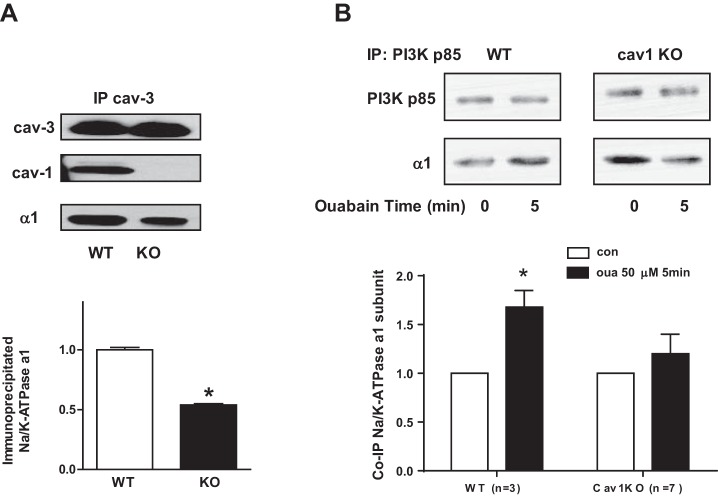
Fig. 7.
Association of Na+/K+-ATPase with signaling proteins in cardiomyocytes from WT and cav-1 KO mice. A: cardiomyocyte lysates were IP with cav-3 antibody and co-IP with cav-1 and Na+/K+-ATPase α1-subunit. Top: representative blots. Bottom: the quantitative data (n = 5). *P < 0.05 vs WT. B: cardiomyocytes were exposed to 50 μM ouabain for 5 min. Cell lysates were subjected to IP with PI3K p85 antibody. Top: representative blots. Bottom: the quantitative data. *P < 0.05 vs. con.

