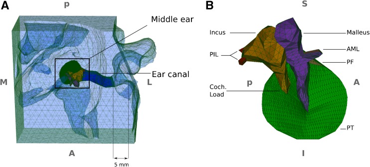
FIG. 1.
Meshed geometry of the finite-element model. A Superior-to-inferior view of the overall model including the ear canal, surrounding soft tissue and middle ear. The 5-mm distance indicates the estimated insertion depth of the probe tip into the canal in the clinical measurements. B Expanded medial-to-lateral view of the middle-ear model, with the TM annulus almost parallel to the page. PIL posterior incudal ligament, AML anterior mallear ligament, PT pars tensa, PF pars flaccida, S superior, I inferior, M medial, L lateral, A anterior, P posterior.

