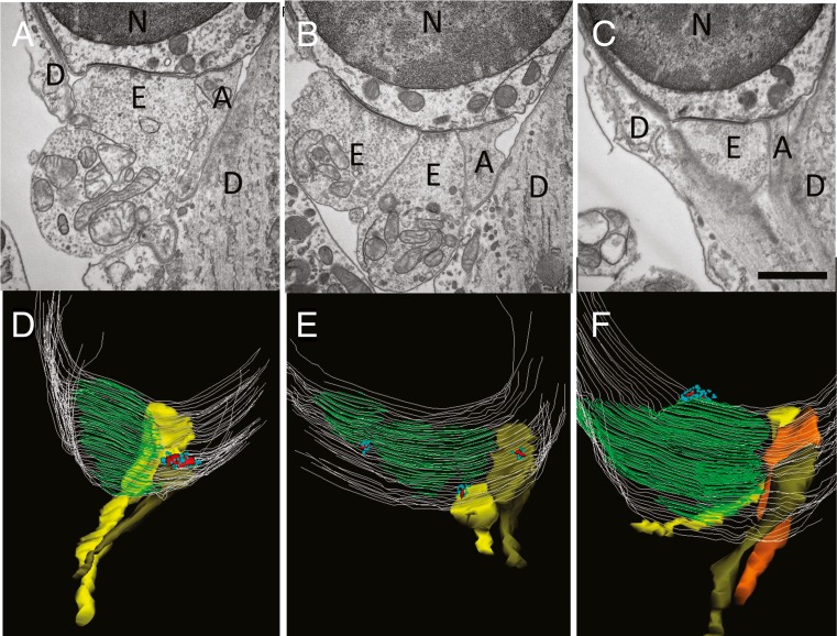
FIG. 7.
Transmission electron micrographs and reconstructions of OHC synapses. A–C Single thin (65 nm) sections from three different OHCs (basal turn, FVB/NJ mouse, P21). Scale bar = 1 μm. Large vesiculated efferent terminals cover most of the synaptic pole and lie opposite to a postsynaptic cistern seen at this magnification as a thickening of the hair cell plasma membrane. Smaller non-vesiculated, granular afferents are opposite hair cell ribbons in approximately half the cases. D–F Z-axis projections of 3D reconstructions. A–C provide example single sections from D–F reconstructions. Hair cell membrane in light gray lines, postsynaptic cisterns colored green. Afferent terminals in yellow, olive, or orange. Synaptic ribbons colored red, nearby vesicles turquoise. Sometimes ribbons appear “misplaced,” e.g., lying over the postsynaptic efferent cistern. Abbreviations N nucleus of outer hair cell, E efferent, A afferent, D Deiters’ cell.

