
Figure 1.
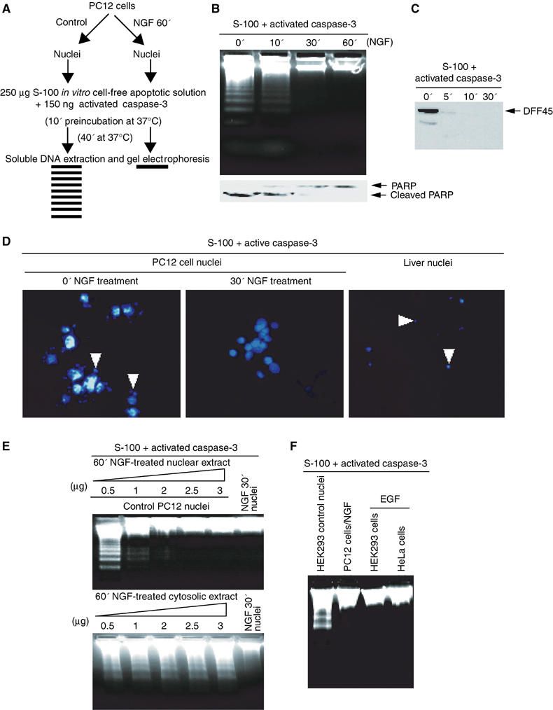
The nuclei from NGF-treated PC12 cells resist DNA fragmentation in cell-free apoptotic solution. (A) Experimental procedures employed in the DNA fragmentation assay. (B) DNA fragmentation assay. The nuclei were isolated from PC12 cells, which were treated with 50 ng/ml NGF (0, 10, 30 min and 1 h) and incubated in the activated cell-free apoptotic solution for 40 min. The fragmented DNA was extracted and resolved on 2% agarose. (C) DFF45 from the control nucleus was cleaved by active caspase-3 in HEK293 cytosolic fraction S-100. (D) Nuclear morphology assay with DAPI staining. The nuclei were isolated from PC12 cells treated with NGF and incubated in apoptotic solution for 40 min and stained with DAPI. The rat liver nuclei were employed as a positive control. (E) PC12 cells were treated with NGF in regular medium for 30 min, and the nuclear and cytosolic extracts of PC12 cells were prepared. Different amounts of the extracts were preincubated with the apoptotic solution for 10 min, then the control nuclei were introduced for DNA fragmentation assay. Nuclear, but not cytosolic, fraction inhibits active apoptosome-triggered DNA fragmentation. (F) The nuclei from HEK293 and HeLa cells, treated with 100 μg/ml EGF for 15 min, are resistant to DNA fragmentation in cell-free apoptotic solution. By contrast, evident DNA fragmentation is observed in control HEK293 cell nuclei without EGF treatment. The nuclei from PC12 cells treated with NGF were employed as positive control.