
Figure 2.
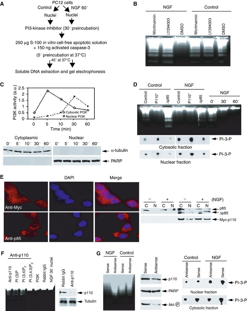
Nuclear PI3K is implicated in NGF-regulated cell survival. (A) Experimental procedures employed in the DNA fragmentation assay. (B) PI3K inhibitors abolish the antiapoptotic effect of NGF in the nucleus. PC12 cells were treated with NGF for 60 min. The isolated nuclei were pre-incubated with two specific PI3K inhibitors Wortmannin (20 nM), Ly294002 (10 μM), and vehicle solution, respectively, for 30 min. After removal of the inhibitors, the nuclei were incubated in apoptotic solution at 37°C for 40 min. The soluble DNA were extracted and analyzed. (C) Cytosolic and nuclear PI3K activity in NGF-treated PC12 cells (upper panel). The identity of cytosolic and nuclear fractions was verified by immunoblotting with anti-α-tubulin and anti-PARP (lower panels). (D) Nuclear PI3K is sufficient and necessary for the antiapoptotic effect of NGF in the nucleus. PC12 cells were infected with control adenovirus and adenovirus expressing constitutively active or dominant-negative PI3K. After 24 h, the infection efficiency was verified by GFP expression under fluorescent microscope. The isolated nuclei from NGF-treated or nontreated PC12 cells were analyzed in apoptotic solution (upper panel). In vitro PI3K activity assay of cytosolic and nuclear fractions from adenovirus-infected PC12 cells (lower panels). (E) PI3K nuclear translocation in PC12 cells infected with adenovirus expressing Myc-p110* and dominant-negative p85. The infected cells were treated with NGF for 30 min, followed by fixation and staining with anti-Myc and anti-p85 antibodies, respectively. Evident nuclear translocation occurred for p110 and p85 proteins (left panels). Similar effects were observed in biochemical fractionations (right panels). (F) Immunodepletion of PI3K from the nuclear extract abolishes its antiapoptotic effect. NGF-treated nuclear extract (10 μg) was preincubated with 2 μl anti-p110 antibody/20 μl protein-A/G conjugated beads at 4°C for 3 h, and the supernatant was supplemented with various phosphoinositol lipids or recombinant PI3K. Evident DNA fragmentation occurs when PI3K was immunodepleted (lane 1), whereas it is potently inhibited when recombinant PI3K was added back (lane 5). By contrast, rabbit IgG control failed to impair the activity (lane 6). Moreover, introduction of 10 μM PI (3,4,5)P3 but not PI (3)P or PI (3,4)P2 reconstitutes the inhibitory effect (left panel). P110 is specifically removed from nuclear extract by anti-p110 antibody (right panels). (G) Nuclear PI3K is required for the antiapoptotic effect of NGF in the nucleus. Serum-starved PC12 cells were treated with Penetratin 1-conjugated sense or antisense oligonucleotides of p110 α for 6 h, followed by 30 min NGF treatment. The isolated nuclei were analyzed in activated apoptotic solution (left panel). The protein level of p110 α and Akt phosphorylation status were verified by Western blotting. Compared to sense oligonucleotide, antisense markedly diminishes p110 α expression. By contrast, PARP protein level is not affected. Akt phosphorylation is substantially decreased in p110-knocked down cells (middle panels). Cytosolic and nuclear PI3K activity is decreased in p110-knocked down PC12 cells (right panels).