
Figure 3.
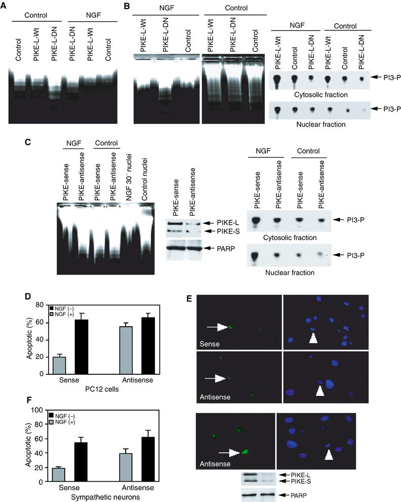
PIKE regulates the antiapoptotic effect of NGF in the nucleus. (A) Dominant-negative PIKE abrogates the antiapoptotic effect of NGF. PC12 cells were infected with control adenovirus and adenovirus expressing wild-type or dominant-negative PIKE. After 24 h, the infection efficiency was verified by GFP expression under a fluorescent microscope. The isolated nuclei from NGF-treated or nontreated PC12 cells were analyzed in apoptotic solution. (B) The stably transfected PC12 cells were induced to express wild-type or dominant-negative PIKE for 24 h, and GFP expression was verified under fluorescent microscope, then followed by NGF or vehicle solution stimulation for 1 h. The isolated nuclei were analyzed in activated apoptotic solution (left panel). Dominant-negative PIKE inhibits cytosolic and nuclear PI3K from induced PIKE stable cell lines (right panels). (C) Serum-starved PC12 cells were treated with Penetratin 1-conjugated sense or antisense oligonucleotides of PIKE for 6 h, followed by 1 h NGF treatment. The isolated nuclei were analyzed in activated cell-free apoptotic solution (left panel). Compared to sense oligonucleotide, antisense markedly diminishes both PIKE-L and -S protein expression. By contrast, PARP protein level is not affected (middle panels). In vitro PI3K activity assay of cytosolic and nuclear PI3K from oligonucleotide-treated cells. PIKE knockdown diminishes NGF-provoked PI3K activity in both the cytoplasm and the nucleus (right panels). (D) PIKE mediates the antiapoptotic effect of NGF in PC12 cells. PC12 cells were treated with Penetratin 1-conjugated sense or antisense oligonucleotides of PIKE for 6 h and induced apoptosis by 250 nM staurosporine for 24 h. (E) TUNEL assay and DAPI staining of staurosporine-treated cells. (F) PIKE mediates the antiapoptotic effect of NGF in sympathetic neurons. Sympathetic neurons were treated with Penetratin 1-conjugated sense or antisense oligonucleotides of PIKE for 6 h and treated with 250 nM staurosporine for 24 h in the presence or absence of NGF. TUNEL assay and DAPI staining of staurosporine-treated apoptotic sympathetic neurons (500 cells were counted under different fields) (right upper panels). Both PIKE-L and -S were markedly knocked down, whereas Tubulin was not changed (right lower panels). Numbers of treated cells in apoptosis were calculated as means (±s.d.) of five determinations and are representative of three experiments.