
Figure 5.
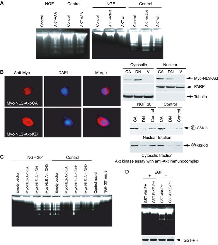
Nuclear Akt is required for the antiapoptotic action of NGF. (A) PC12 cells were infected with control adenovirus and adenovirus expressing dominant-negative Akt (left panel) or wild-type, constitutively active Akt (right panel). After 24 h, the infection efficiency was verified by GFP expression under a fluorescent microscope. The isolated nuclei from PC12 cells were analyzed in an activated cell-free apoptotic solution. (B) Characterization of stably transfected Akt in Myc-NLS-Akt cells. Stably transfected PC12 cells were cultured in tetracycline-free medium overnight, followed by fixation and staining with anti-Myc antibody. Constitutively active (CA)-NLS-Akt predominantly exists in the nucleus, whereas dominant-negative (DN)-NLS-Akt occurs in both the cytoplasm and the nucleus (left panels). Biochemical fractionation and immunoblotting analysis verify the subcellular localization of transfected Akt (right, upper panels). In vitro Akt kinase assay with anti-Akt immunocomplex from the cytosolic and nuclear fractions of PC12 cells, treated with or without NGF (right, lower panels). (C) Stably transfected PC12 cells were induced to express constitutively active or dominant-negative Akt (DN1 and DN2) for 24 h, and GFP expression was verified under a fluorescent microscope, then followed by NGF or vehicle solution stimulation for 30 min. The isolated nuclei were analyzed in the activated apoptotic solution. (D) Expression of PH domains does not interfere with the antiapoptotic actions of EGF. HEK293 cells were transfected with mammalian expression GST-Akt-PH or GST-PIKE-PH, and treated with or without EGF for 15 min. The isolated nuclei were analyzed with DNA fragmentation assay (upper panel). The expression of transfected constructs was verified (lower panel).