
Figure 3.
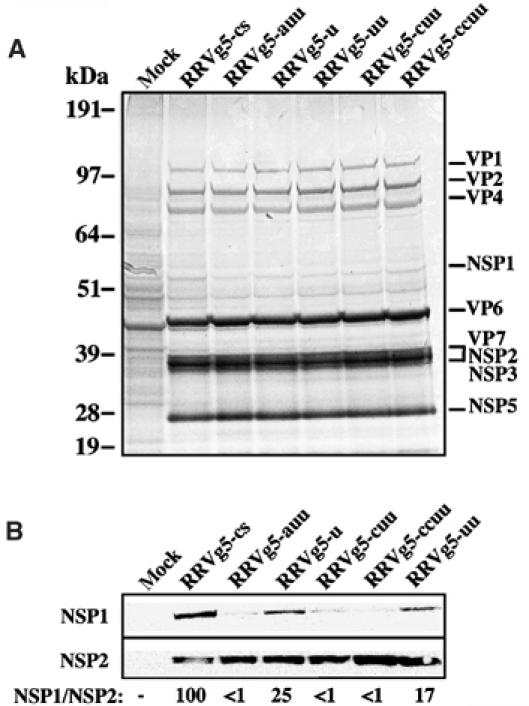
Expression of NSP1 by RRV variants. (A) Lysates prepared from MA104 mock- or RRV-infected MA104 cells, maintained in 35S-amino acids, were analyzed by electrophoresis and autoradiography. NSP1 detection is obscured by a co-migrating host protein. (B) NSP1 and NSP2 in the lysates were detected by Western blot assay with NSP2–NSP1(RRV-C19) antisera. The chemiluminescence exposure shown for NSP2 is for a shorter time than that for NSP1. Intensity values obtained with a phosphorimager were used to calculate the relative ratio of NSP1:NSP2, with the value determined for the RRVg5-cs-infected cells arbitrarily defined as 100. The g5 3′-sequences of the variants are as presented in Table II.