
Figure 2.
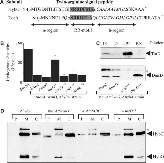
Signal-swapping implicates TorD as a signal peptide chaperone. (A) Amino-acid sequences of the twin-arginine signal peptides from the E. coli hydrogenase-2 β-subunit (HybO) and the TMAO reductase (TorA). The twin-arginine motifs are boxed, the hydrophobic h-regions are shown in italics, and the signal peptidase-I cleavage sites are indicated by the arrows. (B) Hydrogenase-2 activities in mutant strains. Strains RJ606 (‘ΔhybA'), RJ607 φtorA∷hybO, ΔhybA, that produces a HybO protein bearing the TorA signal peptide, and RJ607-D (‘ΔtorD') were grown anaerobically in CR medium containing glycerol and fumarate. In addition, RJ607 was transformed with the pSU series of plasmids that constitutively overproduce TorD (‘torD++'), DmsD (‘dmsD++'), YcdY (‘ycdY++'), or HybE (‘hybE++'), and are grown under identical conditions. Washed whole cells were assayed for hydrogen∷BV oxidoreductase activity with units as μmol BV reduced/min/g cells. (C) Western analysis of TorD and DmsD. Strains RJ607 (φtorA∷hybO, ΔhybA), RJ607-D (‘ΔtorD'), and RJ607 transformed with plasmids overexpressing either torD (‘torD++') or dmsD (‘dmsD++') were cultured anaerobically in CR medium supplemented with glycerol and fumarate. Cells were harvested and resuspended to a concentration of 100 mg (wet weight)/ml (‘1 × '), and a sample diluted to 10 mg/ml (‘10 × '). Identical volumes of protein samples were separated by SDS–PAGE (14% w/v acrylamide), blotted, and challenged with either anti-TorD (top panel) or anti-DmsD (bottom panel) serum at 1:10 000 dilution. (D) Western analysis of the hydrogenase-2 αβ-dimer HybOC. Strains RJ606 (‘ΔhybA'), RJ607 (‘φtorA∷hybO'), RJ607-T (φtorA∷hybO, ΔhybA, ΔtatABC∷KanR; ‘+ ΔtatABC') and RJ607 following transformation with plasmid pSU-torD overexpressing torD (labelled ‘+ torD++') were cultured anaerobically in the presence of glycerol and fumarate. Cells were fractionated in periplasm (P), total membranes (M), and cytoplasm (C), proteins separated by SDS–PAGE (14% w/v acrylamide), blotted, and challenged with an anti-hydrogenase-2 serum. The location of the hydrogenase-2 α-subunit (HybC) and β-subunit (HybO) are indicated. The asterisks denotes a nonspecific immunoreactive band.