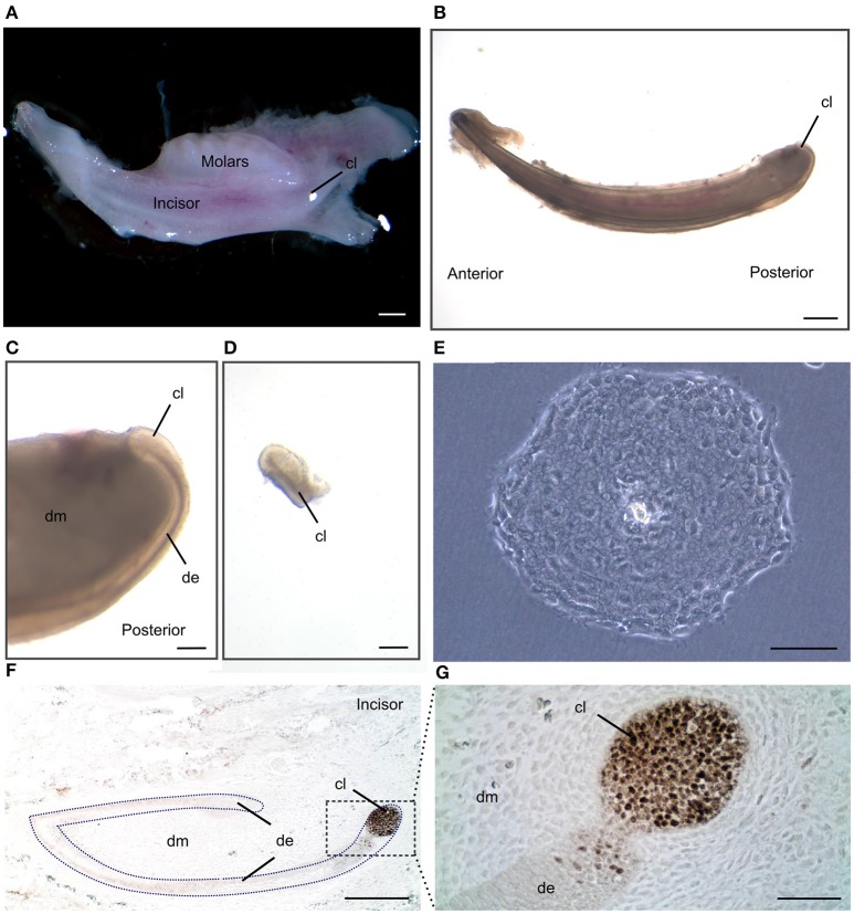
Figure 1.
Isolation of dental epithelial stem cells from mouse incisor cervical loop. (A) Mouse mandibular alveolar bone containing incisor and molars. (B) Isolated postanal mouse incisor. (C) Detail of the posterior part of the incisor where the cervical loop is located. (D) Isolated cervical loop. (E) Colony derived from a single dental epithelial stem cell from mouse cervical loop cultured in a 2D culture system. (F) Immunohistochemistry against the dental epithelial stem cell marker Sox2 and (G) a higher magnification of the cervical loop area. Scale bars: 500 μm in (A,B); 100 μm in (C,D); 200 μm in (E,F); 20 μm in (G).

