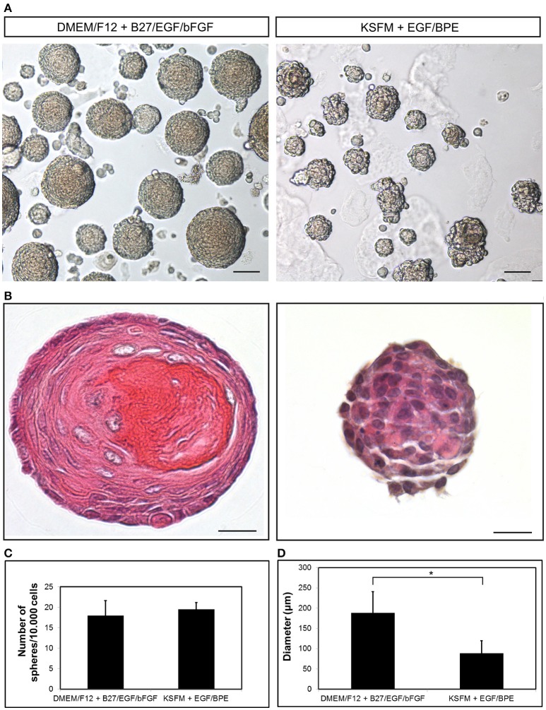
Figure 2.
Growth of dental epithelial stem cells as spheres. (A) Epithelium-derived dentospheres were formed in presence of two different culture media. (B) Hematoxylin-Eosin staining of the resulting spheres. Graphs showing the (C) number and (D) diameter of the spheres. Scale bars: 50 μm in (A); 20 μm in (B). *p < 0.05.

