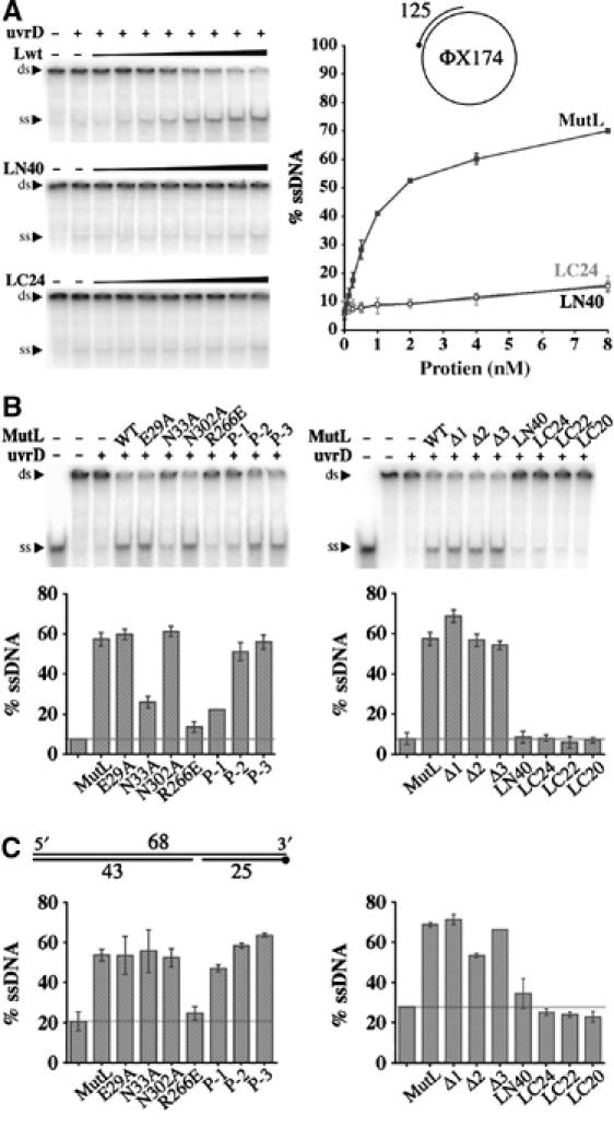
Figure 6.

UvrD activation by MutL. (A) Unwinding of 1 nM circular DNA as illustrated in the cartoon by 1 nM UvrD helicase and increasing amount of MutL, LN40 or LC24 is shown on 4.5% TBE polyacrylamide gels (left panels). The amounts of DNA unwound are quantified and plotted on the right panel. (B) Activation of UvrD by mutant MutL proteins to unwind the circular DNA substrate. Both TBE gels and corresponding bar graphs are shown. (C) Activation of UvrD to unwind a linear DNA substrate as diagrammed. The 5′ end of a 32P-labeled strand is marked by a dot. Only the bar graphs are shown. In each bar graph, the left-most column is UvrD (1 nM) alone, to its right 1 nM of wild-type or mutant MutL is added as labeled. A horizontal line marks the amount of DNA unwound by UvrD alone.
