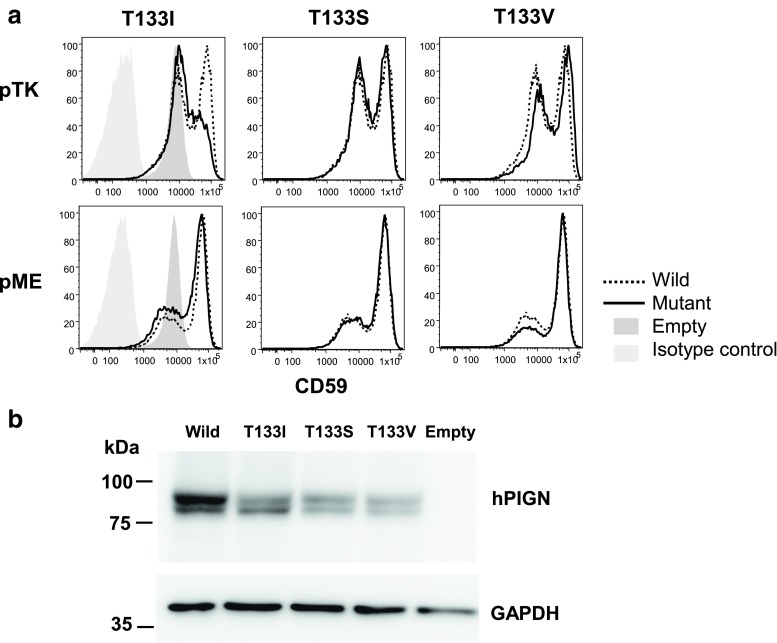
Fig. 2.
Functional analysis of PIGN mutants in PIGN-knockout HEK293 cells. a Restoration of cell-surface expression of GPI-anchored protein CD59 on PIGN-knockout HEK293 cells after transfection. HA-epitope-tagged, wild-type T331I, T331S, and T331V human PIGN cDNAs were transfected with a medium promoter-driven pTK vector (top) or a strong promoter-driven pME vector (bottom). Three days later, CD59 levels were assessed by flow cytometry. b Western blotting analysis of wild-type T331I, T331S, and T331V human PIGN. PIGN-knockout HEK293 cells that were transfected with HA-epitope-tagged, wild-type and mutant PIGN cDNAs in pME were analyzed 3 days later by western blotting using anti-HA antibody. GAPDH glyceraldehyde 3-phosphate dehydrogenase, a loading control

