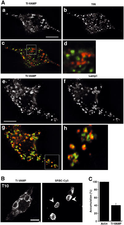
Figure 1.

Endogenous TI-VAMP/VAMP7, colocalised with the lysosomal marker Lamp1, is recruited early and accumulates at the site of phagocytosis. (A) RAW264.7 cells were fixed, permeabilised and stained with anti-TI-VAMP and Cy3-anti-mouse IgG (a, c, e, g) and either anti-TfR (b, d) or anti-Lamp1 (f, h) followed by Cy2-anti-rat IgG. (c, g) Combined images; (d, h) insets in (c, g). Cells were analysed by wide-field fluorescence microscopy with deconvolution. Medial optical sections are shown. Bar, 5 μm. An average of 3.7±0.5 (n=10) TI-VAMP-positive and TfR-positive structures were colocalised per cell representing 8.7±1.1% of total TfR-positive structures while an average of 12±1.6 (n=10) TI-VAMP-positive structures were found to overlap with Lamp1-positive structures per cell which corresponded to 41±5.8% of total Lamp1-positive structures. (B) RAW264.7 cells were incubated for 10 min at 37°C with IgG-SRBCs, and then fixed and stained with Cy3-anti-rabbit IgG (right panel). This staining reveals particles that are still accessible to antibodies and therefore in nonclosed phagosomes, whereas internal particles were observed by phase contrast. The cells were then permeabilised, labelled with anti-TI-VAMP followed by Cy2-anti-mouse IgG (left panel) and analysed by confocal microscopy. One optical section is shown. The arrowheads point to external particles positive for TI-VAMP. Bar, 5 μm. (C) GFP-TI-VAMP is recruited early to phagosomes. RAW264.7 cells transiently transfected to express GFP-TI-VAMP were incubated for 10 min at 37°C with IgG-SRBCs, then placed on ice and, without fixation, stained with Cy3-anti-rabbit IgG to detect external particles. Then, cells were fixed, permeabilised and polymerised actin was labelled with phalloïdine-Alexa350. The number of accumulations of polymerised actin and GFP-TI-VAMP were scored for 50 GFP-TI-VAMP-expressing cells and expressed as an index of accumulation per cell. Then, the index obtained for TI-VAMP recruitment was expressed as a percentage of the index of actin cups. Data are the mean±s.e.m. of three independent experiments.
