
Figure 2.
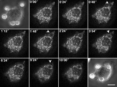
TI-VAMP/VAMP7-positive compartments are recruited early during phagocytosis. RAW264.7 cells transfected to express GFP-TI-VAMP were put into contact with IgG-SRBCs and recorded at 37°C using 4D deconvolution video microscopy. A time-stack was built with maximum intensity projections of deconvolved image stacks (see Movie 1, Supplementary data). Selected images are shown (top left corner, time in s).