
Figure 3.
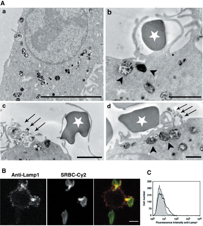
TI-VAMP/Lamp1-positive compartments are exocytosed early during phagocytosis. (A) RAW264.7 cells were allowed to internalise HRP (50 mg/ml) for 30 min at 37°C. After a 1 h chase, phagocytosis was performed for 10 min. The cells were then fixed and processed for DAB cytochemistry and conventional electron microscopy. The arrowheads point to HRP-loaded vesicles recruited to sites of red blood cell attachment and the arrows to exocytosed DAB-positive membranes. The white stars label external SRBCs. Bar, 2 μm. (B) RAW264.7 cells were incubated with IgG-SRBCs for 10 min at 37°C, then placed on ice and, without fixation, stained with anti-Lamp1 antibodies followed by Cy3-anti-rat IgG. External red blood cells were detected with Cy2-anti-rabbit IgG antibodies. Finally, the cells were fixed and analysed by confocal microscopy. One medial optical section is shown. Bar, 5 μm. (C) RAW264.7 cells were treated as in (B), except that secondary antibodies were RPE-coupled. Live cells were then analysed by flow cytometry (bold line). As a control, the cells were incubated with IgG-SRBCs on ice before labelling (dotted histogram).