
Figure 4.
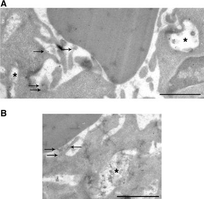
Exocytosis of Lamp1- and TI-VAMP-positive compartments. (A) RAW264.7 macrophages were allowed to phagocytose IgG-SRBCs for 10 min at 37°C and then fixed and processed for ultrathin cryosectioning. Cryosections were immunogold labelled with anti-Lamp1 antibody followed by Protein A-Gold. Bar, 1 μm. Staining of SRBCs was due to a nonspecific reaction. The asterisks indicate Lamp1-positive compartments. The arrows show Lamp1 that was found on membrane ruffles under particles, facing the extracellular medium. (B) Cells were treated as described in (A). Cryosections were immunogold labelled with anti-TI-VAMP antibody followed by Protein A-Gold. Bar, 1 μm. A TI-VAMP-positive compartment is indicated by an asterisk and the arrows show TI-VAMP facing the extracellular medium.