Figure 8.

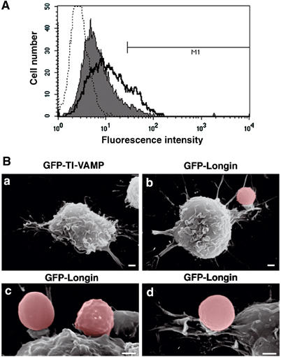
TI-VAMP controls exocytosis and membrane extension during phagocytosis. (A) RAW264.7 cells transfected to express GFP-TI-VAMP or GFP-Longin were incubated with IgG-SRBCs for 10 min at 37°C, then placed on ice and, without fixation, stained with anti-Lamp1 antibodies followed by RPE-anti-rat IgG. At least 5000 live, GFP-positive cells were gated and analysed by flow cytometry. Control cells incubated with secondary antibodies alone (dotted histogram), GFP-Longin-expressing cells (filled histogram), GFP-TI-VAMP-expressing cells (bold line) are shown. M1: Lamp1-positive cells, 18% for GFP-TI-VAMP- and 6% for GFP-Longin-expressing cells. (B) RAW264.7 cells expressing GFP-TI-VAMP (a) or GFP-Longin (b–d) were grown on CeLLocate coverslips, incubated with IgG-SRBCs for 60 min at 37°C, fixed, located on coverslips by fluorescence microscopy and then imaged by SEM. Red blood cells were artificially coloured under Adobe Photoshop 7.0. Bar, 1 μm.
