Figure 3.
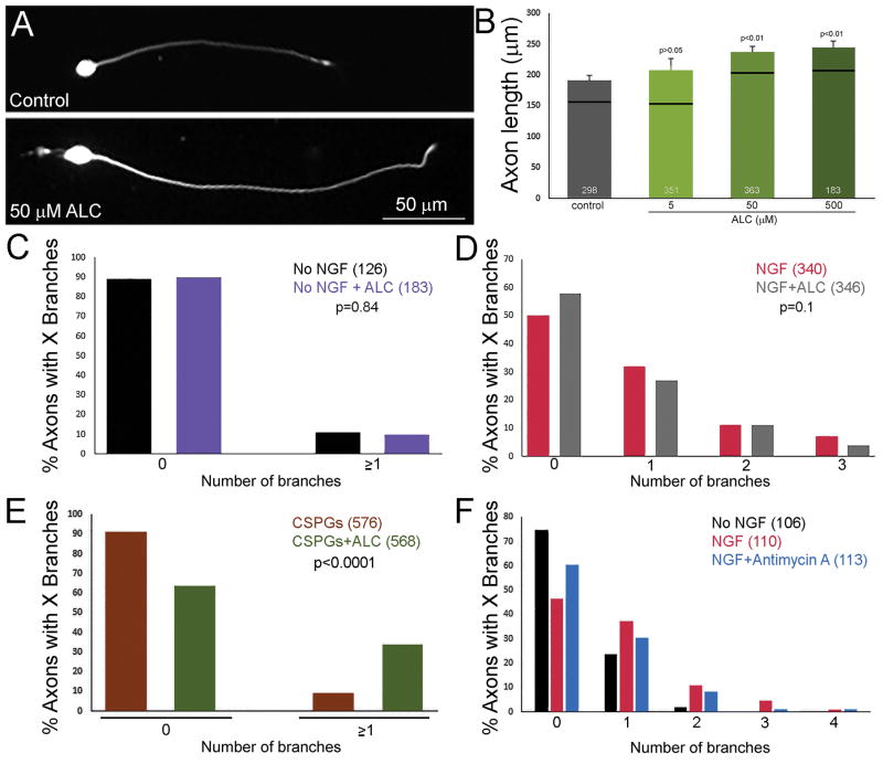
Effects of treatment with ALC on axon development on control and CSPG coated substrata. (A) Examples of dissociated neurons cultured overnight on control substrata in the absence of NGF either treated with 500 μM ALC or control medium. As reflected in the quantification in panel (B), ALC treatment increased axon lengths. (B) Dose dependent effects of ALC treatment on axon lengths on control substrata in the absence of NGF. Black lines represent medians and the number of neurons measured is shown in the bars. n = axons. (C) Graph showing the percentage of axons with 0 or ≥1 branches along their distal 100 μm cultured on control substrata in the absence of NGF ± treatment with 500 μM ALC. n = axons. (D) Graph showing the percentage of axons with 0 to 3 branches along their distal 100 μm cultured on control substrata in the presence of NGF ± treatment with 500 μM ALC. n = axons. (E) Graph showing the percentage of axons with 0 or ≥1 branches along their distal 100 μm cultured on CSPG coated substrata in the presence of NGF ± treatment with 500 μM ALC. n = axons. (F) Graph showing the percentage of axons with 0 to 3 branches along their distal 100 μm cultured overnight on control substrata in the absence of NGF, then treated with NGF for 40 min ± treatment with 2.5 μM antimycin A. NGF induces branching which is attenuated by antimycin A, as reflected by a leftward shit in the distribution. However, the effects of antimycin A was not statistically significant. n = axons.

