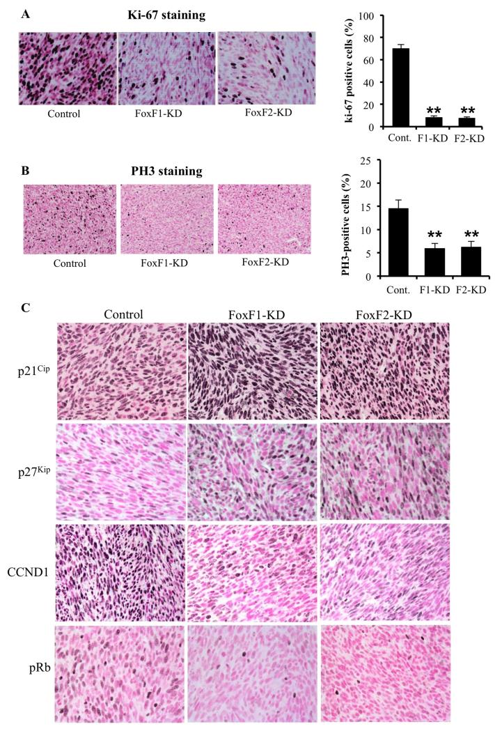
Figure 3. Depletion of FoxF1 and FoxF2 increased expression of p21Cip1 and p27Kip1 in RMS tumors.
RMS tumors were harvested 3 weeks after inoculation of control 76-9 rhabdomyosarcoma cells or 76-9 cells with stable knockdown of FoxF1 or FoxF2 (FoxF1-KD, FoxF2-KD). (A-B) Decreased cellular proliferation was demonstrated by reduced numbers of Ki-67-positive (A) and pH3-positive cells (B). Percentage of Ki-67-positive and PH3-positive cells were counted in five random microscope fields (n=3 mice per group, right panels). A p value < 0.01 is shown with (**). Magnification is 400x (upper panels), 200x (bottom panels). (C) Depletion of FoxF1 or FoxF2 increased expression of p21Cip1 and p27Kip1 CDK inhibitors and decreased protein levels of CCND1 and pRb in RMS tumors. Magnification is 200x.

