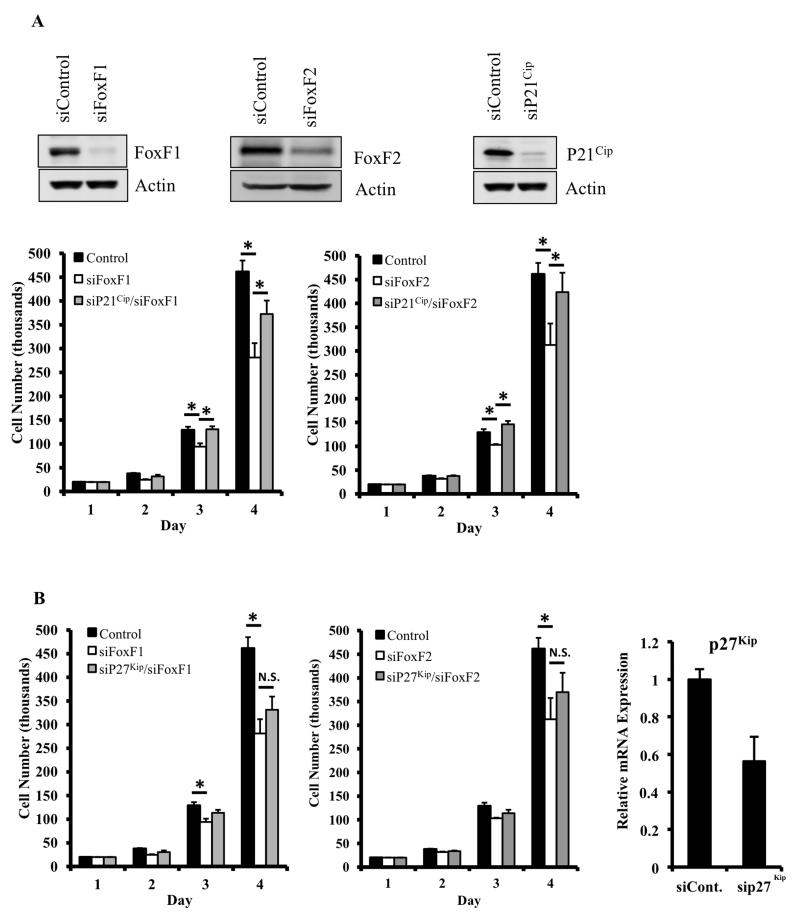
Figure 8. Knockdown of p21Cip1, but not p27Kip1, restores cell proliferation in FoxF1- and FoxF2-deficient cells in vitro.
(A) Western blots showing protein expression FoxF1 (top left), FoxF2 (top middle panel) or p21Cip1 (top right panel) of 91U cells after transfection with control siRNA or siRNA against FoxF1, FoxF2, or p21Cip1. Cell proliferation in the transfected cells was determined by daily cell counts using a hemacytometer. Knockdown of FoxF1 (bottom left) or FoxF2 (bottom right) decreased cell proliferation. Knockdown of p21Cip1 rescued cellular proliferation in both FoxF1- and FoxF2-deficient cells. Data represents the mean ± SD of triplicate wells. (B) Knockdown of p27Kip1 did not restore decreased cellular proliferation in FoxF1- or FoxF2-deficient cells (left and middle panels). Efficiency of p27Kip1 knockdown is shown by qRT-PCR (right panel). Expression levels were normalized to β-actin mRNA. A p value <0.05 is shown with (*).

