
Figure 3.
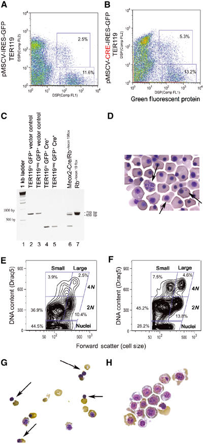
Acute inactivation of pRb in cultured erythroblasts. FACS sort profile of E12.5 FL erythroblasts harvested from Rbflox/flox mice, infected in vitro with control pMSCV-IRES-GFP retrovirus (A) or with Cre recombinase-expressing pMSCV-Cre-IRES-GFP (B) and sorted as GFP-positive/TER119neg- or GFP-positive/TER119lo-expressing cells. PCR analysis (C) of DNA harvested from sorted GFP-positive/TER119neg- (lanes 3 and 5) or GFP-positive/TER119lo- (lanes 2 and 4) expressing cells from either control virus-infected (lanes 2 and 3) or Cre-expressing virus-infected cells (lanes 4 and 5). Excision of floxed exon 19 reduces the size of the Rb PCR product in Rbflox/flox erythroblasts from 700 to 260 bp in erythroblasts infected with Cre-expressing retrovirus (lanes 4 and 5) but not with control retrovirus (lanes 2 and 3). PCR analysis of DNA from E13.5 Rb−/flox FL carrying an Rb null allele (650 bp) and a floxed exon 19 allele (700 bp) (lane 7) and following in vivo excision of the floxed exon 19 on the Meox2-Cre transgenic background generates a 260 bp fragment (lane 6). Peripheral red blood cells from Meox2-Cre/Rb−/exon 19flox E13.5 embryos show abnormal nuclear structure and reduced enucleation (D, arrows). FACS profile of cultured TER119lo/GFP+ erythroblasts following Cre-mediated excision of exon 19 (F) or control cultures (E) labeled with Draq5. Cytospin analysis of TER119lo/GFP+ erythroblasts following 2 days in culture showing extensive red cell enucleation (G, arrows) in the control cultures but not in the Cre-expressing cultures (H).