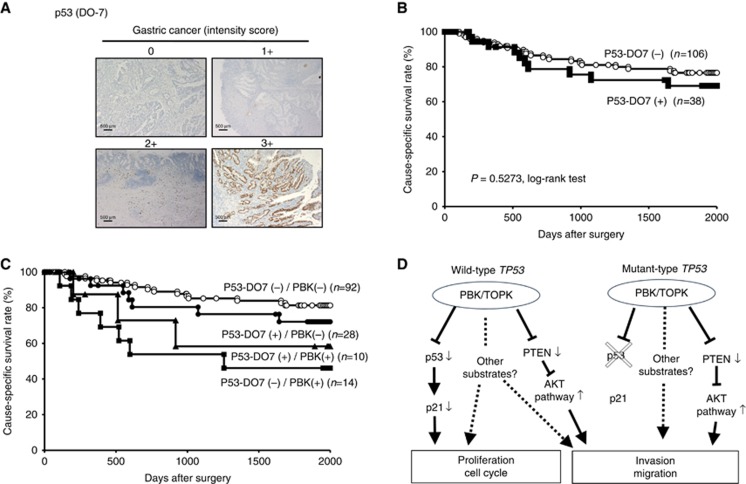
Figure 4.
Relationship between protein expression of PBK/TOPK and p53 in primary cases of gastric cancer. (A) Specific immunostaining of PBK/TOPK in a representative primary tumour sample. On the basis of this result, the intensity scores for p53 (DO7) staining were determined as follows: 0=negative, 1=weak, 2=moderate, 3=strong. Magnification: × 40; Scale bar, 500 μm. (B) Kaplan–Meier curves for cause-specific survival rates of patients at all stages according to the expression of p53. There was no difference of survival between the patients positive and negative for p53-DO7. (C) Postoperative overall survival curves according to a combination of the expression of PBK/TOPK and p53. A significant difference in survival was observed among the patients positive (+) or negative (−) for p53-DO7 and PBK/TOPK (P=0.0228, log-rank test). Notably, in the p53-DO7-negative group, the patients with PBK/TOPK expression had a markedly worse outcome than those with no PBK/TOPK expression. (D) Hypothetical model of the overexpression/activation of PBK/TOPK in GC cells.

