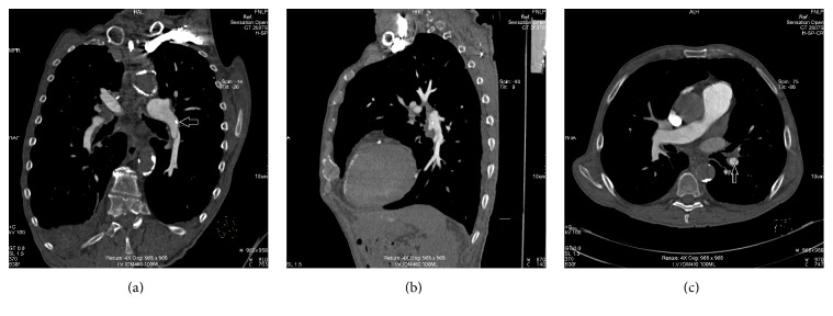
Figure 1.
Coronal and sagittal curved MPR section (a, b) of a contrast-enhanced CT pulmonary angiogram showing long thin dissection flap in the lower lobe branch of the left pulmonary artery with partial involvement of the left pulmonary artery with a small intimal flap calcification (white arrow) and axial section with partial involvement of the lower segmental branch (white arrow) (c).

